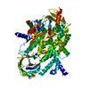
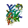
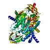
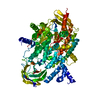
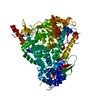
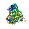
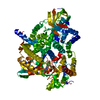
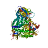
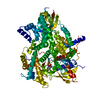
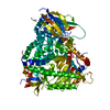
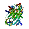
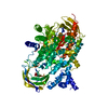
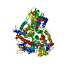
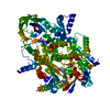
[English] 日本語
Yorodumi
- PDB-7jwe: Gedatolisib bound to the PI3Kg catalytic subunit p110 gamma -

+
Open data


ID or keywords:

Loading...

-
Basic information

Entry
Database: PDB / ID: 7jwe
TitleGedatolisib bound to the PI3Kg catalytic subunit p110 gamma
ComponentsPhosphatidylinositol 4,5-bisphosphate 3-kinase catalytic subunit gamma isoform
KeywordsTRANSFERASE / PIK3CG / mTOR / phosphoinositide / PIP3 / PI3K / p110 / SIGNALING PROTEIN
Function / homology
Function and homology information


secretory granule localization / negative regulation of triglyceride catabolic process / natural killer cell chemotaxis / neutrophil extravasation / phosphatidylinositol-4-phosphate 3-kinase / regulation of calcium ion transmembrane transport / positive regulation of acute inflammatory response / respiratory burst involved in defense response / negative regulation of cardiac muscle contraction / T cell chemotaxis ...secretory granule localization / negative regulation of triglyceride catabolic process / natural killer cell chemotaxis / neutrophil extravasation / phosphatidylinositol-4-phosphate 3-kinase / regulation of calcium ion transmembrane transport / positive regulation of acute inflammatory response / respiratory burst involved in defense response / negative regulation of cardiac muscle contraction / T cell chemotaxis / negative regulation of fibroblast apoptotic process / phosphatidylinositol 3-kinase complex, class IB / sphingosine-1-phosphate receptor signaling pathway / dendritic cell chemotaxis / 1-phosphatidylinositol-4-phosphate 3-kinase activity / 1-phosphatidylinositol-4,5-bisphosphate 3-kinase activity / phosphatidylinositol-4,5-bisphosphate 3-kinase / phosphatidylinositol 3-kinase complex, class IA / phosphatidylinositol 3-kinase / phosphatidylinositol-3-phosphate biosynthetic process / 1-phosphatidylinositol-3-kinase activity / mast cell degranulation / Erythropoietin activates Phosphoinositide-3-kinase (PI3K) / hepatocyte apoptotic process / positive regulation of Rac protein signal transduction / regulation of cell adhesion mediated by integrin / Synthesis of PIPs at the plasma membrane / phosphatidylinositol phosphate biosynthetic process / regulation of angiogenesis / T cell proliferation / cellular response to cAMP / GPVI-mediated activation cascade / ephrin receptor binding / T cell activation / neutrophil chemotaxis / positive regulation of endothelial cell migration / phosphatidylinositol 3-kinase/protein kinase B signal transduction / positive regulation of cytokine production / positive regulation of MAP kinase activity / platelet aggregation / endocytosis / G beta:gamma signalling through PI3Kgamma / Signaling by CSF1 (M-CSF) in myeloid cells / kinase activity / positive regulation of cytosolic calcium ion concentration / angiogenesis / adaptive immune response / positive regulation of phosphatidylinositol 3-kinase/protein kinase B signal transduction / non-specific serine/threonine protein kinase / protein kinase activity / immune response / inflammatory response / G protein-coupled receptor signaling pathway / phosphorylation / protein serine kinase activity / innate immune response / protein serine/threonine kinase activity / ATP binding / membrane / identical protein binding / plasma membrane / cytosol / cytoplasm
Similarity search - Function
PIK3 catalytic subunit gamma, adaptor-binding domain / PIK3 catalytic subunit gamma adaptor-binding domain / Phosphatidylinositol 3-kinase, adaptor-binding domain / Phosphatidylinositol 3-kinase adaptor-binding (PI3K ABD) domain profile. / PI3-kinase family, Ras-binding domain / Phosphatidylinositol 3-kinase Ras-binding (PI3K RBD) domain / PI3-kinase family, ras-binding domain / Phosphatidylinositol 3-kinase Ras-binding (PI3K RBD) domain profile. / Phosphoinositide 3-kinase C2 / Phosphoinositide 3-kinase, region postulated to contain C2 domain ...PIK3 catalytic subunit gamma, adaptor-binding domain / PIK3 catalytic subunit gamma adaptor-binding domain / Phosphatidylinositol 3-kinase, adaptor-binding domain / Phosphatidylinositol 3-kinase adaptor-binding (PI3K ABD) domain profile. / PI3-kinase family, Ras-binding domain / Phosphatidylinositol 3-kinase Ras-binding (PI3K RBD) domain / PI3-kinase family, ras-binding domain / Phosphatidylinositol 3-kinase Ras-binding (PI3K RBD) domain profile. / Phosphoinositide 3-kinase C2 / Phosphoinositide 3-kinase, region postulated to contain C2 domain / C2 phosphatidylinositol 3-kinase-type domain / C2 phosphatidylinositol 3-kinase (PI3K)-type domain profile. / Phosphoinositide 3-kinase, accessory (PIK) domain superfamily / Phosphoinositide 3-kinase family, accessory domain (PIK domain) / Phosphoinositide 3-kinase family, accessory domain (PIK domain) / Phosphoinositide 3-kinase, accessory (PIK) domain / Phosphatidylinositol kinase / PIK helical domain profile. / Phosphatidylinositol 3- and 4-kinases signature 1. / Phosphatidylinositol 3/4-kinase, conserved site / Phosphatidylinositol 3- and 4-kinases signature 2. / Phosphatidylinositol 3-/4-kinase, catalytic domain superfamily / Phosphoinositide 3-kinase, catalytic domain / Phosphatidylinositol 3- and 4-kinase / Phosphatidylinositol 3- and 4-kinases catalytic domain profile. / Phosphatidylinositol 3-/4-kinase, catalytic domain / C2 domain superfamily / Armadillo-type fold / Ubiquitin-like domain superfamily / Protein kinase-like domain superfamily
Similarity search - Domain/homology
Gedatolisib / Phosphatidylinositol 4,5-bisphosphate 3-kinase catalytic subunit gamma isoform
Similarity search - Component
Biological speciesHomo sapiens (human)
MethodX-RAY DIFFRACTION / SYNCHROTRON / MOLECULAR REPLACEMENT / Resolution: 2.55 Å
AuthorsBurke, J.E. / Rathinaswamy, M.K. / Harris, N.J.
Funding support Canada, 1items
OrganizationGrant numberCountry
Canadian Institutes of Health Research (CIHR) Canada
CitationJournal: Elife / Year: 2021
Title: Disease related mutations in PI3K gamma disrupt regulatory C-terminal dynamics and reveal a path to selective inhibitors.
Authors: Rathinaswamy, M.K. / Gaieb, Z. / Fleming, K.D. / Borsari, C. / Harris, N.J. / Moeller, B.J. / Wymann, M.P. / Amaro, R.E. / Burke, J.E.
History
DepositionAug 25, 2020Deposition site: RCSB / Processing site: RCSB
Revision 1.0Mar 17, 2021Provider: repository / Type: Initial release
Revision 1.1Oct 18, 2023Group: Data collection / Database references / Refinement description
Category: chem_comp_atom / chem_comp_bond ...chem_comp_atom / chem_comp_bond / database_2 / pdbx_initial_refinement_model
Item: _database_2.pdbx_DOI / _database_2.pdbx_database_accession

-
Structure visualization

Structure viewerMolecule:
MolmilJmol/JSmol

Downloads & links

-
Assembly

Deposited unit
A: Phosphatidylinositol 4,5-bisphosphate 3-kinase catalytic subunit gamma isoform
hetero molecules


Theoretical massNumber of molelcules
Total (without water)111,3432
Polymers110,7271
Non-polymers6161
Water1629
1


  • Idetical with deposited unit
  • defined by author&software
  • Evidence: gel filtration
TypeNameSymmetry operationNumber
identity operation1_555x,y,z1
MethodPISA
Unit cell
Length a, b, c (Å)143.500, 67.620, 106.330
Angle α, β, γ (deg.)90.000, 95.430, 90.000
Int Tables number5
Space group name H-MC121
Symmetry operation#1: x,y,z
#2: -x,y,-z
#3: x+1/2,y+1/2,z
#4: -x+1/2,y+1/2,-z

-
Components

#1: Protein Phosphatidylinositol 4,5-bisphosphate 3-kinase catalytic subunit gamma isoform / PtdIns-3-kinase subunit gamma / Phosphatidylinositol 4 / 5-bisphosphate 3-kinase 110 kDa catalytic ...PtdIns-3-kinase subunit gamma / Phosphatidylinositol 4 / 5-bisphosphate 3-kinase 110 kDa catalytic subunit gamma / p110gamma / Phosphoinositide-3-kinase catalytic gamma polypeptide / Serine/threonine protein kinase PIK3CG / p120-PI3K


Mass: 110727.102 Da / Num. of mol.: 1
Source method: isolated from a genetically manipulated source
Source: (gene. exp.) Homo sapiens (human) / Gene: PIK3CG / Production host: Spodoptera frugiperda (fall armyworm)
References: UniProt: P48736, phosphatidylinositol-4,5-bisphosphate 3-kinase, non-specific serine/threonine protein kinase
#2: Chemical ChemComp-VL1 / Gedatolisib / N-{4-[4,6-bis(morpholin-4-yl)-1,3,5-triazin-2-yl]phenyl}-N'-{4-[4-(dimethylamino)piperidine-1-carbonyl]phenyl}urea / Gedatolisib


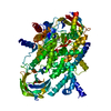
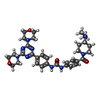
Mass: 615.726 Da / Num. of mol.: 1 / Source method: obtained synthetically / Formula: C32H41N9O4 / Feature type: SUBJECT OF INVESTIGATION
#3: Water ChemComp-HOH / water / Water


Mass: 18.015 Da / Num. of mol.: 9 / Source method: isolated from a natural source / Formula: H2O
Has ligand of interestY

-
Experimental details

-
Experiment

ExperimentMethod: X-RAY DIFFRACTION / Number of used crystals: 1

-
Sample preparation

CrystalDensity Matthews: 2.32 Å3/Da / Density % sol: 46.95 %
Crystal growTemperature: 298 K / Method: vapor diffusion, sitting drop / pH: 7.5
Details: 100 mM Tris pH 7.5, 250 mM (NH4)2SO4 and 20-22% PEG 4000

-
Data collection

DiffractionMean temperature: 77 K / Serial crystal experiment: N
Diffraction sourceSource: SYNCHROTRON / Site: CLSI / Beamline: 08ID-1 / Wavelength: 0.9795 Å
DetectorType: DECTRIS PILATUS3 S 6M / Detector: PIXEL / Date: Aug 24, 2019
RadiationProtocol: SINGLE WAVELENGTH / Monochromatic (M) / Laue (L): M / Scattering type: x-ray
Radiation wavelengthWavelength: 0.9795 Å / Relative weight: 1
ReflectionResolution: 2.55→44.59 Å / Num. obs: 33183 / % possible obs: 99.41 % / Redundancy: 3.3 % / Biso Wilson estimate: 73.57 Å2 / CC1/2: 0.999 / CC star: 0.999 / Rmerge(I) obs: 0.061 / Rpim(I) all: 0.0394 / Rrim(I) all: 0.07285 / Net I/σ(I): 11.91
Reflection shellResolution: 2.55→2.641 Å / Redundancy: 3.4 % / Rmerge(I) obs: 1.349 / Mean I/σ(I) obs: 0.87 / Num. unique obs: 3303 / CC1/2: 0.385 / CC star: 0.746 / Rpim(I) all: 0.8561 / Rrim(I) all: 1.602 / % possible all: 99.4

-
Processing

Software
NameVersionClassification
PHENIX1.18.2_3874refinement
XDSdata reduction
Aimlessdata scaling
PHASERphasing
RefinementMethod to determine structure: MOLECULAR REPLACEMENT
Starting model: 5jha
Resolution: 2.55→44.59 Å / SU ML: 0.48 / Cross valid method: THROUGHOUT / σ(F): 1.35 / Phase error: 31.06 / Stereochemistry target values: ML
RfactorNum. reflection% reflection
Rfree0.2529 1658 5 %
Rwork0.209 31515 -
obs0.2112 33173 99.42 %
Solvent computationShrinkage radii: 0.9 Å / VDW probe radii: 1.11 Å / Solvent model: FLAT BULK SOLVENT MODEL
Displacement parametersBiso max: 187.2 Å2 / Biso mean: 88.8962 Å2 / Biso min: 46.99 Å2
Refinement stepCycle: final / Resolution: 2.55→44.59 Å
ProteinNucleic acidLigandSolventTotal
Num. atoms6612 0 86 9 6707
Biso mean--88.78 65.51 -
Num. residues----815
LS refinement shell

Refine-ID: X-RAY DIFFRACTION / Rfactor Rfree error: 0 / Total num. of bins used: 12

Resolution (Å)Rfactor RfreeNum. reflection RfreeRfactor RworkNum. reflection RworkNum. reflection all% reflection obs (%)
2.55-2.620.45171390.39332630276999
2.62-2.710.36511350.341725952730100
2.71-2.810.34251380.287726082746100
2.81-2.920.32121370.27592599273699
2.92-3.050.31741380.26826332771100
3.05-3.210.33321370.251926042741100
3.21-3.410.26851380.23032612275099
3.41-3.680.27521380.209826142752100
3.68-4.050.25081370.18992622275999
4.05-4.630.18131400.16382646278699
4.63-5.830.21711390.19232641278099
5.83-44.590.24241420.19212711285399
Refinement TLS params.

Method: refined / Refine-ID: X-RAY DIFFRACTION

IDL112)L122)L132)L222)L232)L332)S11 (Å °)S12 (Å °)S13 (Å °)S21 (Å °)S22 (Å °)S23 (Å °)S31 (Å °)S32 (Å °)S33 (Å °)T112)T122)T132)T222)T232)T332)Origin x (Å)Origin y (Å)Origin z (Å)
13.94250.0624-0.38081.1091-0.53572.48290.01340.0538-0.51020.01210.18030.33460.0822-0.09740.00020.72540.0148-0.0290.5026-0.00020.786432.4592-28.020624.3913
23.7849-0.76580.98151.1744-0.05462.24180.0643-0.0219-0.40930.00570.0447-0.03390.10810.46090.00010.68080.00370.02460.5779-0.02540.683551.4597-25.963630.1933
33.8573-1.17840.982.765-0.20251.6434-0.19480.08660.38350.15250.10610.3206-0.4023-0.3722-0.00030.72590.06050.03210.6090.0540.684723.6856-7.680425.2945
40.5965-0.15790.02550.5043-0.46530.4023-0.4247-0.25141.0390.4429-0.1156-0.8175-0.54670.2435-0.00071.36180.0335-0.24750.9002-0.15171.261138.99212.7237.9537
Refinement TLS group
IDRefine-IDRefine TLS-IDSelection detailsAuth asym-IDAuth seq-ID
1X-RAY DIFFRACTION1chain 'A' and (resid 145 through 422 )A145 - 422
2X-RAY DIFFRACTION2chain 'A' and (resid 423 through 688 )A423 - 688
3X-RAY DIFFRACTION3chain 'A' and (resid 689 through 1037 )A689 - 1037
4X-RAY DIFFRACTION4chain 'A' and (resid 1038 through 1090 )A0

+
About Yorodumi

-
News

-
Feb 9, 2022. New format data for meta-information of EMDB entries

New format data for meta-information of EMDB entries

  • Version 3 of the EMDB header file is now the official format.
  • The previous official version 1.9 will be removed from the archive.

Related info.:EMDB header

External links:wwPDB to switch to version 3 of the EMDB data model

-
Aug 12, 2020. Covid-19 info

Covid-19 info

URL: https://pdbj.org/emnavi/covid19.php

New page: Covid-19 featured information page in EM Navigator.

Related info.:Covid-19 info / Mar 5, 2020. Novel coronavirus structure data

+
Mar 5, 2020. Novel coronavirus structure data

Novel coronavirus structure data

Related info.:Yorodumi Speices / Aug 12, 2020. Covid-19 info

External links:COVID-19 featured content - PDBj / Molecule of the Month (242):Coronavirus Proteases

+
Jan 31, 2019. EMDB accession codes are about to change! (news from PDBe EMDB page)

EMDB accession codes are about to change! (news from PDBe EMDB page)

  • The allocation of 4 digits for EMDB accession codes will soon come to an end. Whilst these codes will remain in use, new EMDB accession codes will include an additional digit and will expand incrementally as the available range of codes is exhausted. The current 4-digit format prefixed with “EMD-” (i.e. EMD-XXXX) will advance to a 5-digit format (i.e. EMD-XXXXX), and so on. It is currently estimated that the 4-digit codes will be depleted around Spring 2019, at which point the 5-digit format will come into force.
  • The EM Navigator/Yorodumi systems omit the EMD- prefix.

Related info.:Q: What is EMD? / ID/Accession-code notation in Yorodumi/EM Navigator

External links:EMDB Accession Codes are Changing Soon! / Contact to PDBj

+
Jul 12, 2017. Major update of PDB

Major update of PDB

  • wwPDB released updated PDB data conforming to the new PDBx/mmCIF dictionary.
  • This is a major update changing the version number from 4 to 5, and with Remediation, in which all the entries are updated.
  • In this update, many items about electron microscopy experimental information are reorganized (e.g. em_software).
  • Now, EM Navigator and Yorodumi are based on the updated data.

External links:wwPDB Remediation / Enriched Model Files Conforming to OneDep Data Standards Now Available in the PDB FTP Archive

-
Yorodumi

Thousand views of thousand structures

  • Yorodumi is a browser for structure data from EMDB, PDB, SASBDB, etc.
  • This page is also the successor to EM Navigator detail page, and also detail information page/front-end page for Omokage search.
  • The word "yorodu" (or yorozu) is an old Japanese word meaning "ten thousand". "mi" (miru) is to see.

Related info.:EMDB / PDB / SASBDB / Comparison of 3 databanks / Yorodumi Search / Aug 31, 2016. New EM Navigator & Yorodumi / Yorodumi Papers / Jmol/JSmol / Function and homology information / Changes in new EM Navigator and Yorodumi

Read more