 Movie
Movie Controller

[English] 日本語
 Yorodumi
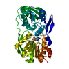
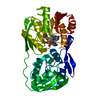
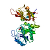
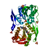
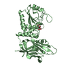
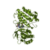
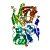
- PDB-7jvx: Crystal structure of PTEN (aa 7-353 followed by spacer TGGGSGGTGG... -
+ Open data

- Basic information
| Entry | Database: PDB / ID: 7jvx | |||||||||
|---|---|---|---|---|---|---|---|---|---|---|
| Title | Crystal structure of PTEN (aa 7-353 followed by spacer TGGGSGGTGGGSGGTGGGCY ligated to peptide pSDpTpTDpSDPENEPFDED) | |||||||||
 Components Components | Phosphatidylinositol 3,4,5-trisphosphate 3-phosphatase and dual-specificity protein phosphatase PTEN | |||||||||
 Keywords Keywords |  SIGNALING PROTEIN / phosphatase / Phosphatidylinositol 3 / 4 / 5-trisphosphate 3-phosphatase and dual-specificity protein phosphatase PTEN SIGNALING PROTEIN / phosphatase / Phosphatidylinositol 3 / 4 / 5-trisphosphate 3-phosphatase and dual-specificity protein phosphatase PTEN | |||||||||
| Function / homology |  Function and homology information Function and homology informationinositol-1,3,4,5,6-pentakisphosphate 3-phosphatase activity / PTEN Loss of Function in Cancer / phosphatidylinositol-3,4,5-trisphosphate 3-phosphatase / regulation of cellular component size / negative regulation of keratinocyte migration / phosphatidylinositol-3,4-bisphosphate 3-phosphatase activity / negative regulation of synaptic vesicle clustering / phosphatidylinositol phosphate phosphatase activity / rhythmic synaptic transmission / inositol-1,3,4,5-tetrakisphosphate 3-phosphatase activity ...inositol-1,3,4,5,6-pentakisphosphate 3-phosphatase activity / PTEN Loss of Function in Cancer / phosphatidylinositol-3,4,5-trisphosphate 3-phosphatase / regulation of cellular component size / negative regulation of keratinocyte migration / phosphatidylinositol-3,4-bisphosphate 3-phosphatase activity / negative regulation of synaptic vesicle clustering / phosphatidylinositol phosphate phosphatase activity / rhythmic synaptic transmission / inositol-1,3,4,5-tetrakisphosphate 3-phosphatase activity / central nervous system myelin maintenance / negative regulation of wound healing, spreading of epidermal cells / phosphatidylinositol-3-phosphate phosphatase activity / central nervous system neuron axonogenesis / postsynaptic density assembly / neuron-neuron synaptic transmission / negative regulation of dendritic spine morphogenesis / presynaptic membrane assembly / Regulation of PTEN mRNA translation / synapse maturation / Negative regulation of the PI3K/AKT network / cellular response to electrical stimulus / negative regulation of cell cycle G1/S phase transition / negative regulation of axonogenesis / Transcriptional Regulation by MECP2 / phosphatidylinositol-3,4,5-trisphosphate 3-phosphatase activity / myelin sheath adaxonal region / negative regulation of excitatory postsynaptic potential / negative regulation of organ growth / forebrain morphogenesis / negative regulation of focal adhesion assembly / phosphatidylinositol dephosphorylation / Schmidt-Lanterman incisure / anaphase-promoting complex binding / negative regulation of epithelial to mesenchymal transition / Hydrolases; Acting on ester bonds; Phosphoric-monoester hydrolases / dentate gyrus development / negative regulation of cyclin-dependent protein serine/threonine kinase activity / positive regulation of ubiquitin protein ligase activity / spindle assembly involved in female meiosis / positive regulation of ubiquitin-dependent protein catabolic process / phosphatidylinositol biosynthetic process / negative regulation of cell size / dendritic spine morphogenesis / brain morphogenesis / myosin phosphatase activity / protein serine/threonine phosphatase activity / negative regulation of G1/S transition of mitotic cell cycle / ubiquitin-specific protease binding / molecular function inhibitor activity / protein-serine/threonine phosphatase / regulation of neuron projection development / regulation of phosphatidylinositol 3-kinase/protein kinase B signal transduction / Synthesis of IP3 and IP4 in the cytosol / locomotor rhythm / phosphoprotein phosphatase activity / negative regulation of vascular associated smooth muscle cell proliferation / negative regulation of cellular senescence / social behavior / Synthesis of PIPs at the plasma membrane / positive regulation of excitatory postsynaptic potential / negative regulation of phosphatidylinositol 3-kinase/protein kinase B signal transduction / canonical Wnt signaling pathway / multicellular organismal response to stress / localization / prepulse inhibition / negative regulation of peptidyl-serine phosphorylation / synapse assembly / Regulation of PTEN localization / protein dephosphorylation / protein-tyrosine-phosphatase / negative regulation of cell migration / negative regulation of protein phosphorylation / locomotory behavior / phosphatidylinositol 3-kinase/protein kinase B signal transduction / central nervous system development / protein tyrosine phosphatase activity / cell projection / cell motility / PDZ domain binding / TP53 Regulates Metabolic Genes / regulation of protein stability / cytoplasmic side of plasma membrane / PML body / Regulation of PTEN stability and activity / positive regulation of DNA-binding transcription factor activity / cell migration / Ovarian tumor domain proteases / Downstream TCR signaling / negative regulation of neuron projection development / heart development / dendritic spine / postsynaptic density / learning or memory / protein stabilization / Ub-specific processing proteases / neuron projection / apical plasma membrane / negative regulation of cell population proliferation / apoptotic processSimilarity search - Function | |||||||||
| Biological species |  Homo sapiens (human) Homo sapiens (human) | |||||||||
| Method | X-RAY DIFFRACTION / SYNCHROTRON / MOLECULAR REPLACEMENT / Resolution: 3.2 Å | |||||||||
| Model details | Crystal Structure of 4p-PTEN (aa 7-353+ 20 aa spacer, GGGSGGTGGGSGGTGGGCY+CRYpSDpTpTDpSDPENEG | |||||||||
 Authors Authors | Dempsey, D. / Phan, K. / Cole, P. / Gabelli, S.B. | |||||||||
| Funding support |  United States, 2items United States, 2items
| |||||||||
 Citation Citation | Journal: Nat.Struct.Mol.Biol. / Year: 2021 Title: The structural basis of PTEN regulation by multi-site phosphorylation. Authors: Dempsey, D.R. / Viennet, T. / Iwase, R. / Park, E. / Henriquez, S. / Chen, Z. / Jeliazkov, J.R. / Palanski, B.A. / Phan, K.L. / Coote, P. / Gray, J.J. / Eck, M.J. / Gabelli, S.B. / Arthanari, H. / Cole, P.A. | |||||||||
| History |
|
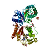
- Structure visualization
| Structure viewer | Molecule: MolmilJmol/JSmol |
|---|
- Downloads & links
-Download
| PDBx/mmCIF format | 7jvx.cif.gz | 82.4 KB | Display | PDBx/mmCIF format |
|---|---|---|---|---|
| PDB format | pdb7jvx.ent.gz | 59.4 KB | Display | PDB format |
| PDBx/mmJSON format | 7jvx.json.gz | Tree view | PDBx/mmJSON format | |
| Others |  Other downloads Other downloads |
-Validation report
| Arichive directory | https://data.pdbj.org/pub/pdb/validation_reports/jv/7jvxftp://data.pdbj.org/pub/pdb/validation_reports/jv/7jvx | HTTPS FTP |
|---|
-Related structure data
| Related structure data | 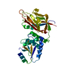 7jtxC 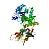 7jukC 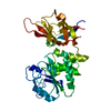 7julC  1d5rS S: Starting model for refinement C: citing same article ( |
|---|---|
| Similar structure data |

-Links
 PDBj
PDBj


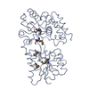
- Assembly
| Deposited unit | 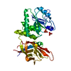
| ||||||||
|---|---|---|---|---|---|---|---|---|---|
| 1 |
| ||||||||
| Unit cell |
|
-Components
| #1: Protein | Mass: 48555.484 Da / Num. of mol.: 1 Source method: isolated from a genetically manipulated source Source: (gene. exp.) Homo sapiens (human) / Gene: PTEN, MMAC1, TEP1 / Plasmid: pfastbac / Production host:  Trichoplusia ni (cabbage looper) Trichoplusia ni (cabbage looper)References: UniProt: P60484, protein-serine/threonine phosphatase, protein-tyrosine-phosphatase, phosphatidylinositol-3,4,5-trisphosphate 3-phosphatase | ||
|---|---|---|---|
| #2: Chemical | Phosphate  Mass: 94.971 Da / Num. of mol.: 2 / Source method: obtained synthetically / Formula: PO4 Mass: 94.971 Da / Num. of mol.: 2 / Source method: obtained synthetically / Formula: PO4Has ligand of interest | N | |
-Experimental details
-Experiment
| Experiment | Method: X-RAY DIFFRACTION / Number of used crystals: 1 |
|---|
- Sample preparation
| Crystal | Density Matthews: 1.91 Å3/Da / Density % sol: 35.49 % |
|---|---|
| Crystal grow | Temperature: 293 K / Method: vapor diffusion / Details: 300 mM citrate |
-Data collection
| Diffraction | Mean temperature: 277 K / Serial crystal experiment: N | ||||||||||||||||||||||||||||||||||||||||||||||||||||||||||||||||||||||||||||||||
|---|---|---|---|---|---|---|---|---|---|---|---|---|---|---|---|---|---|---|---|---|---|---|---|---|---|---|---|---|---|---|---|---|---|---|---|---|---|---|---|---|---|---|---|---|---|---|---|---|---|---|---|---|---|---|---|---|---|---|---|---|---|---|---|---|---|---|---|---|---|---|---|---|---|---|---|---|---|---|---|---|---|
| Diffraction source | Source: SYNCHROTRON / Site: NSLS-II / Beamline: 17-ID-2 / Wavelength: 0.9791 Å | ||||||||||||||||||||||||||||||||||||||||||||||||||||||||||||||||||||||||||||||||
| Detector | Type: DECTRIS EIGER X 16M / Detector: PIXEL / Date: Nov 3, 2017 | ||||||||||||||||||||||||||||||||||||||||||||||||||||||||||||||||||||||||||||||||
| Radiation | Protocol: SINGLE WAVELENGTH / Monochromatic (M) / Laue (L): M / Scattering type: x-ray | ||||||||||||||||||||||||||||||||||||||||||||||||||||||||||||||||||||||||||||||||
| Radiation wavelength | Wavelength: 0.9791 Å / Relative weight: 1 | ||||||||||||||||||||||||||||||||||||||||||||||||||||||||||||||||||||||||||||||||
| Reflection | Resolution: 2.9→19.8 Å / Num. obs: 7482 / % possible obs: 90.3 % / Redundancy: 2.691 % / Biso Wilson estimate: 68.088 Å2 / CC1/2: 0.973 / Rmerge(I) obs: 0.212 / Rrim(I) all: 0.252 / Χ2: 0.883 / Net I/σ(I): 3.19 / Num. measured all: 20136 / Scaling rejects: 8 | ||||||||||||||||||||||||||||||||||||||||||||||||||||||||||||||||||||||||||||||||
| Reflection shell | Diffraction-ID: 1
|
- Processing
| Software |
| ||||||||||||||||||||||||||||||||||||||||||||||||||||||||||||
|---|---|---|---|---|---|---|---|---|---|---|---|---|---|---|---|---|---|---|---|---|---|---|---|---|---|---|---|---|---|---|---|---|---|---|---|---|---|---|---|---|---|---|---|---|---|---|---|---|---|---|---|---|---|---|---|---|---|---|---|---|---|
| Refinement | Method to determine structure: MOLECULAR REPLACEMENT Starting model: 1D5R Resolution: 3.2→19.8 Å / Cor.coef. Fo:Fc: 0.92 / Cor.coef. Fo:Fc free: 0.862 / WRfactor Rfree: 0.2944 / WRfactor Rwork: 0.2509 / FOM work R set: 0.6512 / SU B: 51.656 / SU ML: 0.812 / SU Rfree: 0.6801 / Cross valid method: THROUGHOUT / σ(F): 0 / ESU R Free: 0.68 / Stereochemistry target values: MAXIMUM LIKELIHOOD Details: HYDROGENS HAVE BEEN ADDED IN THE RIDING POSITIONS U VALUES : REFINED INDIVIDUALLY
| ||||||||||||||||||||||||||||||||||||||||||||||||||||||||||||
| Solvent computation | Ion probe radii: 0.8 Å / Shrinkage radii: 0.8 Å / VDW probe radii: 1.2 Å / Solvent model: MASK | ||||||||||||||||||||||||||||||||||||||||||||||||||||||||||||
| Displacement parameters | Biso max: 182.8 Å2 / Biso mean: 85.335 Å2 / Biso min: 30 Å2
| ||||||||||||||||||||||||||||||||||||||||||||||||||||||||||||
| Refinement step | Cycle: final / Resolution: 3.2→19.8 Å
| ||||||||||||||||||||||||||||||||||||||||||||||||||||||||||||
| Refine LS restraints |
| ||||||||||||||||||||||||||||||||||||||||||||||||||||||||||||
| LS refinement shell | Resolution: 3.2→3.283 Å / Rfactor Rfree error: 0 / Total num. of bins used: 20
|
