 Movie
Movie Controller


+ Open data

- Basic information
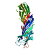
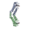
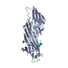
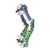
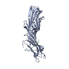
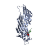
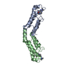
| Entry | Database: PDB / ID: 7jof | ||||||
|---|---|---|---|---|---|---|---|
| Title | Calcium-bound C2A Domain from Human Dysferlin | ||||||
 Components Components | Isoform 6 of Dysferlin | ||||||
 Keywords Keywords |  MEMBRANE PROTEIN / Membrane repair / calcium-binding / C2 domain / phospholipid binding MEMBRANE PROTEIN / Membrane repair / calcium-binding / C2 domain / phospholipid binding | ||||||
| Function / homology |  Function and homology information Function and homology informationmonocyte activation involved in immune response / regulation of neurotransmitter secretion / macrophage activation involved in immune response / calcium-dependent phospholipid binding / negative regulation of phagocytosis / centriolar satellite / endocytic vesicle / Smooth Muscle Contraction / T-tubule / cytoplasmic vesicle membrane ...monocyte activation involved in immune response / regulation of neurotransmitter secretion / macrophage activation involved in immune response / calcium-dependent phospholipid binding / negative regulation of phagocytosis / centriolar satellite / endocytic vesicle / Smooth Muscle Contraction / T-tubule / cytoplasmic vesicle membrane / phospholipid binding / sarcolemma / synaptic vesicle membrane / late endosome / early endosome / endosome / calcium ion binding / extracellular exosome / plasma membraneSimilarity search - Function | ||||||
| Biological species |  Homo sapiens (human) Homo sapiens (human) | ||||||
| Method | X-RAY DIFFRACTION / MOLECULAR REPLACEMENT / Resolution: 2 Å | ||||||
 Authors Authors | Tadayon, R. / Wang, Y. / Santamaria, L. / Mercier, P. / Forristal, C. / Shaw, G.S. | ||||||
| Funding support |  Canada, 1items Canada, 1items
| ||||||
 Citation Citation | Journal: Biochem.J. / Year: 2021 Title: Calcium binds and rigidifies the dysferlin C2A domain in a tightly coupled manner. Authors: Wang, Y. / Tadayon, R. / Santamaria, L. / Mercier, P. / Forristal, C.J. / Shaw, G.S. | ||||||
| History |
|
- Structure visualization
| Structure viewer | Molecule: MolmilJmol/JSmol |
|---|
- Downloads & links
-Download
| PDBx/mmCIF format | 7jof.cif.gz | 119.4 KB | Display | PDBx/mmCIF format |
|---|---|---|---|---|
| PDB format | pdb7jof.ent.gz | 90.1 KB | Display | PDB format |
| PDBx/mmJSON format | 7jof.json.gz | Tree view | PDBx/mmJSON format | |
| Others |  Other downloads Other downloads |
-Validation report
| Arichive directory | https://data.pdbj.org/pub/pdb/validation_reports/jo/7jofftp://data.pdbj.org/pub/pdb/validation_reports/jo/7jof | HTTPS FTP |
|---|
-Related structure data
| Related structure data |  7k6bC 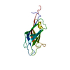 7krbC 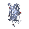 4ihbS S: Starting model for refinement C: citing same article ( |
|---|---|
| Similar structure data |

-Links
 PDBj
PDBj


- Assembly
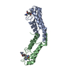
| Deposited unit | 
| ||||||||
|---|---|---|---|---|---|---|---|---|---|
| 1 | 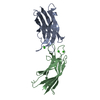
| ||||||||
| 2 | 
| ||||||||
| Unit cell |
|
-Components
| #1: Protein | Mass: 14765.858 Da / Num. of mol.: 4 Source method: isolated from a genetically manipulated source Source: (gene. exp.) Homo sapiens (human) / Gene: DYSF, FER1L1 / Plasmid: pET28a / Production host:  Escherichia coli BL21(DE3) (bacteria) / References: UniProt: O75923 Escherichia coli BL21(DE3) (bacteria) / References: UniProt: O75923#2: Chemical | ChemComp-CA /   Mass: 40.078 Da / Num. of mol.: 8 / Source method: obtained synthetically / Formula: Ca / Feature type: SUBJECT OF INVESTIGATION Mass: 40.078 Da / Num. of mol.: 8 / Source method: obtained synthetically / Formula: Ca / Feature type: SUBJECT OF INVESTIGATION#3: Water | ChemComp-HOH / | Water Mass: 18.015 Da / Num. of mol.: 378 / Source method: isolated from a natural source / Formula: H2O Mass: 18.015 Da / Num. of mol.: 378 / Source method: isolated from a natural source / Formula: H2OHas ligand of interest | Y | |
|---|
-Experimental details
-Experiment
| Experiment | Method: X-RAY DIFFRACTION / Number of used crystals: 1 |
|---|
- Sample preparation
| Crystal | Density Matthews: 2.06 Å3/Da / Density % sol: 40.27 % |
|---|---|
| Crystal grow | Temperature: 283.15 K / Method: vapor diffusion, sitting drop / pH: 8.5 Details: 0.2 mM magnesium chloride hexahydrate, 0.1 M Tris hydrochloride, and 30% Polyethylene glycol 4,000 (commercial reagent kit from Hampton Research, HR2-130) at pH 8.5 |
-Data collection
| Diffraction | Mean temperature: 100 K / Serial crystal experiment: N | ||||||||||||||||||||||||||||||
|---|---|---|---|---|---|---|---|---|---|---|---|---|---|---|---|---|---|---|---|---|---|---|---|---|---|---|---|---|---|---|---|
| Diffraction source | Source: ROTATING ANODE / Type: RIGAKU MICROMAX-007 / Wavelength: 1.5 Å | ||||||||||||||||||||||||||||||
| Detector | Type: RIGAKU SATURN 944+ / Detector: CCD / Date: Feb 5, 2017 | ||||||||||||||||||||||||||||||
| Radiation | Protocol: SINGLE WAVELENGTH / Monochromatic (M) / Laue (L): M / Scattering type: x-ray | ||||||||||||||||||||||||||||||
| Radiation wavelength | Wavelength: 1.5 Å / Relative weight: 1 | ||||||||||||||||||||||||||||||
| Reflection | Resolution: 1.99→23.72 Å / Num. obs: 28309 / % possible obs: 87.6 % / Redundancy: 1.7 % / Biso Wilson estimate: 12.26 Å2 / CC1/2: 0.992 / Rmerge(I) obs: 0.061 / Rpim(I) all: 0.061 / Rrim(I) all: 0.086 / Net I/σ(I): 9 / Num. measured all: 47404 | ||||||||||||||||||||||||||||||
| Reflection shell | Diffraction-ID: 1
|
- Processing
| Software |
| ||||||||||||||||||||||||||||||||||||||||||
|---|---|---|---|---|---|---|---|---|---|---|---|---|---|---|---|---|---|---|---|---|---|---|---|---|---|---|---|---|---|---|---|---|---|---|---|---|---|---|---|---|---|---|---|
| Refinement | Method to determine structure: MOLECULAR REPLACEMENT Starting model: 4ihb Resolution: 2→9.986 Å / SU ML: 0.19 / Cross valid method: THROUGHOUT / σ(F): 1.98 / Phase error: 24.86 / Stereochemistry target values: ML
| ||||||||||||||||||||||||||||||||||||||||||
| Solvent computation | Shrinkage radii: 0.9 Å / VDW probe radii: 1.11 Å / Solvent model: FLAT BULK SOLVENT MODEL | ||||||||||||||||||||||||||||||||||||||||||
| Displacement parameters | Biso max: 69.31 Å2 / Biso mean: 17.5728 Å2 / Biso min: 1.68 Å2 | ||||||||||||||||||||||||||||||||||||||||||
| Refinement step | Cycle: final / Resolution: 2→9.986 Å
| ||||||||||||||||||||||||||||||||||||||||||
| LS refinement shell | Refine-ID: X-RAY DIFFRACTION / Rfactor Rfree error: 0
|
