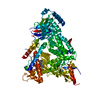
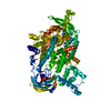
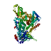
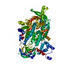
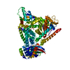
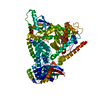
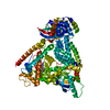
Entry Database : PDB / ID : 7jiuTitle HUMAN PI3KDELTA IN COMPLEX WITH COMPOUND 2F Phosphatidylinositol 4,5-bisphosphate 3-kinase catalytic subunit alpha isoform Keywords / / / Function / homology Function Domain/homology Component
/ / / / / / / / / / / / / / / / / / / / / / / / / / / / / / / / / / / / / / / / / / / / / / / / / / / / / / / / / / / / / / / / / / / / / / / / / / / / / / / / / / / / / / / / / / / / / / / / / / / / / / / / / / / / / / / / / / / / / / / / / / / / / / / / / / / / / / / / / / / / / / / / / / / Biological species Homo sapiens (human)Method / / / Resolution : 2.12 Å Authors Lesburg, C.A. / Augustin, M. Journal : Acs Med.Chem.Lett. / Year : 2020Title : Optimization of Versatile Oxindoles as Selective PI3K delta Inhibitors.Authors : Methot, J.L. / Achab, A. / Christopher, M. / Zhou, H. / McGowan, M.A. / Trotter, B.W. / Fradera, X. / Lesburg, C.A. / Goldenblatt, P. / Hill, A. / Chen, D. / Otte, K.M. / Augustin, M. / Shah, S. / Katz, J.D. History Deposition Jul 23, 2020 Deposition site / Processing site Revision 1.0 Jun 30, 2021 Provider / Type Revision 1.1 Mar 6, 2024 Group / Data collection / Database referencesCategory chem_comp_atom / chem_comp_bond ... chem_comp_atom / chem_comp_bond / database_2 / pdbx_unobs_or_zero_occ_atoms Item / _database_2.pdbx_database_accessionRevision 1.2 Apr 3, 2024 Group / Category
Show all Show less
 Movie
Movie Controller


 Open data
 Basic information
 Components
 Keywords
 TRANSFERASE / TRANSFERASE-TRANSFERASE INHIBITOR COMPLEX
 Function and homology information
 Authors
 Citation
 Structure visualization
 Downloads & links
 Other downloads
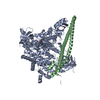


 PDBj
PDBj


 Assembly
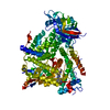

 Mass: 475.544 Da / Num. of mol.: 1 / Source method: obtained synthetically / Formula: C28H25N7O / Feature type: SUBJECT OF INVESTIGATION
 Mass: 18.015 Da / Num. of mol.: 289 / Source method: isolated from a natural source / Formula: H2O
 Sample preparation
 / Beamline: X06SA / Wavelength: 1.00003 Å
 Processing