 Movie
Movie Controller

[English] 日本語
 Yorodumi
- PDB-7jgo: Crystal Structure of the Ni-bound Human Heavy-chain variant 122H-... -
+ Open data

- Basic information
| Entry | Database: PDB / ID: 7jgo | ||||||
|---|---|---|---|---|---|---|---|
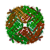
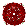
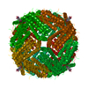
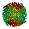
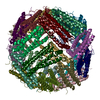
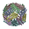
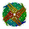
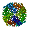
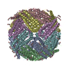
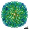
| Title | Crystal Structure of the Ni-bound Human Heavy-chain variant 122H-delta C-star with 2,5-furandihyrdoxamate collected at 278K | ||||||
 Components Components | Ferritin heavy chain | ||||||
 Keywords Keywords | OXIDOREDUCTASE / Protein-MOF / Ferritin-MOF / Self-Assembly / Ferritin | ||||||
| Function / homology |  Function and homology information Function and homology informationiron ion sequestering activity / ferritin complex / Scavenging by Class A Receptors / negative regulation of ferroptosis / Golgi Associated Vesicle Biogenesis / ferroxidase / autolysosome / ferroxidase activity / negative regulation of fibroblast proliferation / ferric iron binding ...iron ion sequestering activity / ferritin complex / Scavenging by Class A Receptors / negative regulation of ferroptosis / Golgi Associated Vesicle Biogenesis / ferroxidase / autolysosome / ferroxidase activity / negative regulation of fibroblast proliferation / ferric iron binding / autophagosome / Iron uptake and transport / iron ion transport / ferrous iron binding / tertiary granule lumen / ficolin-1-rich granule lumen / intracellular iron ion homeostasis / immune response / iron ion binding / negative regulation of cell population proliferation / Neutrophil degranulation / extracellular exosome / extracellular region / identical protein binding / nucleus / cytosol / cytoplasm Similarity search - Function | ||||||
| Biological species |  Homo sapiens (human) Homo sapiens (human) | ||||||
| Method |  X-RAY DIFFRACTION / MOLECULAR REPLACEMENT / Resolution: 3.08 Å X-RAY DIFFRACTION / MOLECULAR REPLACEMENT / Resolution: 3.08 Å | ||||||
 Authors Authors | Bailey, J.B. / Tezcan, F.A. | ||||||
| Funding support |  United States, 1items United States, 1items
| ||||||
 Citation Citation | Journal: J.Am.Chem.Soc. / Year: 2020 Title: Tunable and Cooperative Thermomechanical Properties of Protein-Metal-Organic Frameworks. Authors: Bailey, J.B. / Tezcan, F.A. | ||||||
| History |
|
- Structure visualization
| Structure viewer | Molecule: MolmilJmol/JSmol |
|---|
- Downloads & links
-Download
| PDBx/mmCIF format | 7jgo.cif.gz | 62.3 KB | Display | PDBx/mmCIF format |
|---|---|---|---|---|
| PDB format | pdb7jgo.ent.gz | 36.7 KB | Display | PDB format |
| PDBx/mmJSON format | 7jgo.json.gz | Tree view | PDBx/mmJSON format | |
| Others |  Other downloads Other downloads |
-Validation report
| Summary document | 7jgo_validation.pdf.gz | 432.9 KB | Display | wwPDB validaton report |
|---|---|---|---|---|
| Full document | 7jgo_full_validation.pdf.gz | 433.1 KB | Display | |
| Data in XML | 7jgo_validation.xml.gz | 8.9 KB | Display | |
| Data in CIF | 7jgo_validation.cif.gz | 11.2 KB | Display | |
| Arichive directory | https://data.pdbj.org/pub/pdb/validation_reports/jg/7jgoftp://data.pdbj.org/pub/pdb/validation_reports/jg/7jgo | HTTPS FTP |
-Related structure data
| Related structure data | 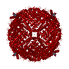 7jgkC  7jglC 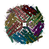 7jgmC 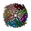 7jgnC 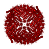 7jgpC 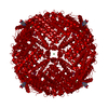 7jgqC 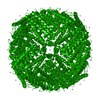 5cmqS S: Starting model for refinement C: citing same article ( |
|---|---|
| Similar structure data |


-Links
 PDBj
PDBj


- Assembly
| Deposited unit | 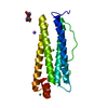
| ||||||||||||||||||||||||
|---|---|---|---|---|---|---|---|---|---|---|---|---|---|---|---|---|---|---|---|---|---|---|---|---|---|
| 1 | 
| ||||||||||||||||||||||||
| Unit cell |
| ||||||||||||||||||||||||
| Components on special symmetry positions |
|
-Components
| #1: Protein | Mass: 21122.291 Da / Num. of mol.: 1 Source method: isolated from a genetically manipulated source Source: (gene. exp.) Homo sapiens (human) / Gene: FTH1, FTH, FTHL6, OK/SW-cl.84, PIG15 / Production host:  | ||||||
|---|---|---|---|---|---|---|---|
| #2: Chemical | ChemComp-V9Y /   Mass: 186.122 Da / Num. of mol.: 1 / Source method: obtained synthetically / Formula: C6H6N2O5 Mass: 186.122 Da / Num. of mol.: 1 / Source method: obtained synthetically / Formula: C6H6N2O5 | ||||||
| #3: Chemical |   Mass: 58.693 Da / Num. of mol.: 3 / Source method: obtained synthetically / Formula: Ni Mass: 58.693 Da / Num. of mol.: 3 / Source method: obtained synthetically / Formula: Ni#4: Chemical | ChemComp-NA / |   Mass: 22.990 Da / Num. of mol.: 1 / Source method: obtained synthetically / Formula: Na Mass: 22.990 Da / Num. of mol.: 1 / Source method: obtained synthetically / Formula: Na#5: Water | ChemComp-HOH / |  Mass: 18.015 Da / Num. of mol.: 25 / Source method: isolated from a natural source / Formula: H2O Mass: 18.015 Da / Num. of mol.: 25 / Source method: isolated from a natural source / Formula: H2OHas ligand of interest | Y | |
-Experimental details
-Experiment
| Experiment | Method: X-RAY DIFFRACTION / Number of used crystals: 1 |
|---|
- Sample preparation
| Crystal | Density Matthews: 3.75 Å3/Da / Density % sol: 67.2 % |
|---|---|
| Crystal grow | Temperature: 298 K / Method: vapor diffusion, sitting drop / pH: 9.5 Details: Reservoir: 500 uL total volume: 50 mM CHES (pH 9.5), 150 mM NaCl, 0.474 mM NiCl2, 12.6% PEP Sitting Drop: 12.7 uL reservoir, 3.3 uL of 25 uM ferritin, 4 uL of 10 mM fdh in 50 mM CHES (pH 9.5) with 150 mM NaCl |
-Data collection
| Diffraction | Mean temperature: 278 K / Serial crystal experiment: N |
|---|---|
| Diffraction source | Source: ROTATING ANODE / Type: BRUKER AXS MICROSTAR / Wavelength: 1.54178 Å |
| Detector | Type: APEX II CCD / Detector: CCD / Date: Sep 6, 2019 |
| Radiation | Protocol: SINGLE WAVELENGTH / Monochromatic (M) / Laue (L): M / Scattering type: x-ray |
| Radiation wavelength | Wavelength: 1.54178 Å / Relative weight: 1 |
| Reflection | Resolution: 3.08→49.36 Å / Num. obs: 11398 / % possible obs: 100 % / Redundancy: 18.4 % / Biso Wilson estimate: 48.66 Å2 / CC1/2: 0.996 / Net I/σ(I): 9.8 |
| Reflection shell | Resolution: 3.08→3.19 Å / Num. unique obs: 1026 / CC1/2: 0.899 |
- Processing
| Software |
| |||||||||||||||||||||||||||||||||||||||||||||||||||||||||||||||
|---|---|---|---|---|---|---|---|---|---|---|---|---|---|---|---|---|---|---|---|---|---|---|---|---|---|---|---|---|---|---|---|---|---|---|---|---|---|---|---|---|---|---|---|---|---|---|---|---|---|---|---|---|---|---|---|---|---|---|---|---|---|---|---|---|
| Refinement | Method to determine structure: MOLECULAR REPLACEMENT Starting model: 5CMQ Resolution: 3.08→49.36 Å / SU ML: 0.359 / Cross valid method: FREE R-VALUE / σ(F): 1.33 / Phase error: 26.3106 Stereochemistry target values: GeoStd + Monomer Library + CDL v1.2
| |||||||||||||||||||||||||||||||||||||||||||||||||||||||||||||||
| Solvent computation | Shrinkage radii: 0.9 Å / VDW probe radii: 1.11 Å / Solvent model: FLAT BULK SOLVENT MODEL | |||||||||||||||||||||||||||||||||||||||||||||||||||||||||||||||
| Displacement parameters | Biso mean: 39.25 Å2 | |||||||||||||||||||||||||||||||||||||||||||||||||||||||||||||||
| Refinement step | Cycle: LAST / Resolution: 3.08→49.36 Å
| |||||||||||||||||||||||||||||||||||||||||||||||||||||||||||||||
| Refine LS restraints |
| |||||||||||||||||||||||||||||||||||||||||||||||||||||||||||||||
| LS refinement shell |
|
