 Movie
Movie Controller

[English] 日本語
 Yorodumi
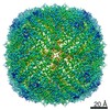
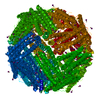
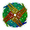
- PDB-5cmr: Crystal Structure of Linker-Mediated Zn-bound Human H-Ferritin va... -
+ Open data

- Basic information
| Entry | Database: PDB / ID: 5cmr | ||||||
|---|---|---|---|---|---|---|---|
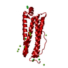
| Title | Crystal Structure of Linker-Mediated Zn-bound Human H-Ferritin variant 122H-delta C-star | ||||||
 Components Components | Ferritin heavy chain | ||||||
 Keywords Keywords | OXIDOREDUCTASE / Protein Engineering / Metal Binding / Supramolecular Assembly | ||||||
| Function / homology |  Function and homology information Function and homology informationiron ion sequestering activity / : / autolysosome / Scavenging by Class A Receptors / Golgi Associated Vesicle Biogenesis / ferroxidase / intracellular sequestering of iron ion / ferroxidase activity / negative regulation of fibroblast proliferation / ferric iron binding ...iron ion sequestering activity / : / autolysosome / Scavenging by Class A Receptors / Golgi Associated Vesicle Biogenesis / ferroxidase / intracellular sequestering of iron ion / ferroxidase activity / negative regulation of fibroblast proliferation / ferric iron binding / ferrous iron binding / Iron uptake and transport / tertiary granule lumen / iron ion transport / intracellular iron ion homeostasis / ficolin-1-rich granule lumen / immune response / iron ion binding / negative regulation of cell population proliferation / Neutrophil degranulation / extracellular exosome / extracellular region / identical protein binding / nucleus / cytosol / cytoplasmSimilarity search - Function | ||||||
| Biological species |  Homo sapiens (human) Homo sapiens (human) | ||||||
| Method | X-RAY DIFFRACTION / SYNCHROTRON / MOLECULAR REPLACEMENT / Resolution: 3.792 Å | ||||||
 Authors Authors | Sontz, P.A. / Bailey, J.B. / Ahn, S. / Tezcan, F.A. | ||||||
| Funding support |  United States, 1items United States, 1items
| ||||||
 Citation Citation | Journal: J.Am.Chem.Soc. / Year: 2015 Title: A Metal Organic Framework with Spherical Protein Nodes: Rational Chemical Design of 3D Protein Crystals. Authors: Sontz, P.A. / Bailey, J.B. / Ahn, S. / Tezcan, F.A. | ||||||
| History |
|
- Structure visualization
| Structure viewer | Molecule: MolmilJmol/JSmol |
|---|
- Downloads & links
-Download
| PDBx/mmCIF format | 5cmr.cif.gz | 53.7 KB | Display | PDBx/mmCIF format |
|---|---|---|---|---|
| PDB format | pdb5cmr.ent.gz | 36.9 KB | Display | PDB format |
| PDBx/mmJSON format | 5cmr.json.gz | Tree view | PDBx/mmJSON format | |
| Others |  Other downloads Other downloads |
-Validation report
| Arichive directory | https://data.pdbj.org/pub/pdb/validation_reports/cm/5cmrftp://data.pdbj.org/pub/pdb/validation_reports/cm/5cmr | HTTPS FTP |
|---|
-Related structure data
| Related structure data |  5cmqC  2ceiS C: citing same article ( S: Starting model for refinement |
|---|---|
| Similar structure data |


-Links
 PDBj
PDBj


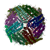
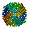
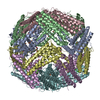
- Assembly
| Deposited unit | 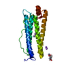
| ||||||||||||||||||
|---|---|---|---|---|---|---|---|---|---|---|---|---|---|---|---|---|---|---|---|
| 1 | x 24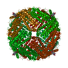
| ||||||||||||||||||
| Unit cell |
| ||||||||||||||||||
| Components on special symmetry positions |
|
-Components
| #1: Protein | / Ferritin H subunit / Cell proliferation-inducing gene 15 protein Mass: 21065.240 Da / Num. of mol.: 1 Fragment: Ferritin-like diiron domain containing residues 6-178 Mutation: K86A, C90E, C102A, C130A, T122H Source method: isolated from a genetically manipulated source Source: (gene. exp.) Homo sapiens (human) / Gene: FTH1, FTH, FTHL6, OK/SW-cl.84, PIG15 / Production host:  Escherichia coli (E. coli) / References: UniProt: P02794, ferroxidase Escherichia coli (E. coli) / References: UniProt: P02794, ferroxidase | ||||
|---|---|---|---|---|---|
| #2: Chemical |   Mass: 65.409 Da / Num. of mol.: 2 / Source method: obtained synthetically / Formula: Zn Mass: 65.409 Da / Num. of mol.: 2 / Source method: obtained synthetically / Formula: Zn#3: Chemical |   Mass: 22.990 Da / Num. of mol.: 3 / Source method: obtained synthetically / Formula: Na Mass: 22.990 Da / Num. of mol.: 3 / Source method: obtained synthetically / Formula: Na#4: Chemical | ChemComp-BYD / |   Mass: 196.160 Da / Num. of mol.: 1 / Source method: obtained synthetically / Formula: C8H8N2O4 Mass: 196.160 Da / Num. of mol.: 1 / Source method: obtained synthetically / Formula: C8H8N2O4 |
-Experimental details
-Experiment
| Experiment | Method: X-RAY DIFFRACTION / Number of used crystals: 1 |
|---|
- Sample preparation
| Crystal | Density Matthews: 3.71 Å3/Da / Density % sol: 66.88 % |
|---|---|
| Crystal grow | Temperature: 298 K / Method: vapor diffusion, sitting drop / pH: 8.5 Details: 50 mM CHES, 150 mM sodium chloride, 0.3 mM zinc chloride, 1 mM H2BDH |
-Data collection
| Diffraction | Mean temperature: 100 K | ||||||||||||||||||||||||||||||||||||||||||||||||||||||||||||||||||||||||||||||||||||||||||||||||||||||||||||||
|---|---|---|---|---|---|---|---|---|---|---|---|---|---|---|---|---|---|---|---|---|---|---|---|---|---|---|---|---|---|---|---|---|---|---|---|---|---|---|---|---|---|---|---|---|---|---|---|---|---|---|---|---|---|---|---|---|---|---|---|---|---|---|---|---|---|---|---|---|---|---|---|---|---|---|---|---|---|---|---|---|---|---|---|---|---|---|---|---|---|---|---|---|---|---|---|---|---|---|---|---|---|---|---|---|---|---|---|---|---|---|---|
| Diffraction source | Source: SYNCHROTRON / Site: SSRL / Beamline: BL12-2 / Wavelength: 0.98 Å | ||||||||||||||||||||||||||||||||||||||||||||||||||||||||||||||||||||||||||||||||||||||||||||||||||||||||||||||
| Detector | Type: DECTRIS PILATUS 6M / Detector: PIXEL / Date: Apr 18, 2015 | ||||||||||||||||||||||||||||||||||||||||||||||||||||||||||||||||||||||||||||||||||||||||||||||||||||||||||||||
| Radiation | Monochromator: Rh coated flat mirror / Protocol: SINGLE WAVELENGTH / Monochromatic (M) / Laue (L): M / Scattering type: x-ray | ||||||||||||||||||||||||||||||||||||||||||||||||||||||||||||||||||||||||||||||||||||||||||||||||||||||||||||||
| Radiation wavelength | Wavelength: 0.98 Å / Relative weight: 1 | ||||||||||||||||||||||||||||||||||||||||||||||||||||||||||||||||||||||||||||||||||||||||||||||||||||||||||||||
| Reflection | Resolution: 3.792→109.828 Å / Num. all: 3401 / Num. obs: 3401 / % possible obs: 99.8 % / Redundancy: 11.2 % / Biso Wilson estimate: 94.54 Å2 / Rpim(I) all: 0.056 / Rrim(I) all: 0.194 / Rsym value: 0.185 / Net I/av σ(I): 4.125 / Net I/σ(I): 12 / Num. measured all: 38132 | ||||||||||||||||||||||||||||||||||||||||||||||||||||||||||||||||||||||||||||||||||||||||||||||||||||||||||||||
| Reflection shell | Diffraction-ID: 1 / Rejects: 0
|
- Processing
| Software |
| ||||||||||||||||||||||||
|---|---|---|---|---|---|---|---|---|---|---|---|---|---|---|---|---|---|---|---|---|---|---|---|---|---|
| Refinement | Method to determine structure: MOLECULAR REPLACEMENT Starting model: 2CEI Resolution: 3.792→54.914 Å / SU ML: 0.4 / Cross valid method: THROUGHOUT / σ(F): 1.37 / Phase error: 22.92 / Stereochemistry target values: ML
| ||||||||||||||||||||||||
| Solvent computation | Shrinkage radii: 0.9 Å / VDW probe radii: 1.11 Å / Solvent model: FLAT BULK SOLVENT MODEL | ||||||||||||||||||||||||
| Displacement parameters | Biso max: 145.88 Å2 / Biso mean: 81.9409 Å2 / Biso min: 47.02 Å2 | ||||||||||||||||||||||||
| Refinement step | Cycle: final / Resolution: 3.792→54.914 Å
| ||||||||||||||||||||||||
| Refine LS restraints |
| ||||||||||||||||||||||||
| LS refinement shell | Refine-ID: X-RAY DIFFRACTION / Total num. of bins used: 2
|
