 Movie
Movie Controller

[English] 日本語
 Yorodumi
- PDB-7fqi: Crystal Structure of human Legumain in complex with (2S)-N-[(1S)-... -
+ Open data

- Basic information
| Entry | Database: PDB / ID: 7fqi | |||||||||
|---|---|---|---|---|---|---|---|---|---|---|
| Title | Crystal Structure of human Legumain in complex with (2S)-N-[(1S)-3-amino-1-cyano-3-oxopropyl]-1-[1-[4-[(2,4-difluorophenyl)methoxy]phenyl]cyclopropanecarbonyl]pyrrolidine-2-carboxamide | |||||||||
 Components Components | Legumain Asparagine endopeptidase Asparagine endopeptidase | |||||||||
 Keywords Keywords | HYDROLASE/INHIBITOR / CYSTEINE PROTEASE / ALLOSTERIC INHIBITOR / ASPARAGINYL ENDOPEPTIDASE / ALZHEIMER'S DISEASE / HYDROLASE / HYDROLASE-INHIBITOR complex | |||||||||
| Function / homology |  Function and homology information Function and homology informationnegative regulation of ERBB signaling pathway / legumain / vacuolar protein processing / renal system process / Vitamin D (calciferol) metabolism / receptor catabolic process / vitamin D metabolic process / self proteolysis / endolysosome lumen / activation of cysteine-type endopeptidase activity ...negative regulation of ERBB signaling pathway / legumain / vacuolar protein processing / renal system process / Vitamin D (calciferol) metabolism / receptor catabolic process / vitamin D metabolic process / self proteolysis / endolysosome lumen / activation of cysteine-type endopeptidase activity / positive regulation of endothelial cell chemotaxis / response to acidic pH / dendritic spine organization / positive regulation of monocyte chemotaxis / Trafficking and processing of endosomal TLR / negative regulation of multicellular organism growth / cellular response to hepatocyte growth factor stimulus / associative learning / protein maturation / endopeptidase activator activity / cellular response to calcium ion / MHC class II antigen presentation / proteolysis involved in protein catabolic process / positive regulation of mitotic cell cycle / lysosomal lumen / positive regulation of long-term synaptic potentiation / tau protein binding / memory / cellular response to amyloid-beta / antigen processing and presentation of exogenous peptide antigen via MHC class II / late endosome / apical part of cell / peptidase activity / negative regulation of neuron apoptotic process / lysosome / cysteine-type endopeptidase activity / negative regulation of gene expression / positive regulation of cell population proliferation / perinuclear region of cytoplasm / proteolysis / extracellular exosome / extracellular region / cytoplasmSimilarity search - Function | |||||||||
| Biological species |  Homo sapiens (human) Homo sapiens (human) | |||||||||
| Method | X-RAY DIFFRACTION / SYNCHROTRON / MOLECULAR REPLACEMENT / Resolution: 1.45 Å | |||||||||
 Authors Authors | Ehler, A. / Benz, J. / Bartels, B. / Rudolph, M.G. | |||||||||
| Funding support |  Switzerland, 1items Switzerland, 1items
| |||||||||
 Citation Citation | Journal: To be published Title: Crystal Structure of a human Legumain complex Authors: Bartels, B. / Kuhn, B. / Benz, J. / Rudolph, M.G. | |||||||||
| History |
|
- Structure visualization
| Structure viewer | Molecule: MolmilJmol/JSmol |
|---|
- Downloads & links
-Download
| PDBx/mmCIF format | 7fqi.cif.gz | 199.3 KB | Display | PDBx/mmCIF format |
|---|---|---|---|---|
| PDB format | pdb7fqi.ent.gz | 153.7 KB | Display | PDB format |
| PDBx/mmJSON format | 7fqi.json.gz | Tree view | PDBx/mmJSON format | |
| Others |  Other downloads Other downloads |
-Validation report
| Arichive directory | https://data.pdbj.org/pub/pdb/validation_reports/fq/7fqiftp://data.pdbj.org/pub/pdb/validation_reports/fq/7fqi | HTTPS FTP |
|---|
-Group deposition
| ID | G_1002245 (5 entries) |
|---|---|
| Title | To be published |
| Type | undefined |
| Description | A set of AEP_Legumain crystal structures |


-Related structure data
| Similar structure data |
|---|
-Links
 PDBj
PDBj


- Assembly
| Deposited unit | 
| ||||||||
|---|---|---|---|---|---|---|---|---|---|
| 1 |
| ||||||||
| 2 |
| ||||||||
| 3 |
| ||||||||
| Unit cell |
|
-Components
| #1: Protein | Asparagine endopeptidase / Asparaginyl endopeptidase / Protease / cysteine 1 Mass: 50877.672 Da / Num. of mol.: 3 Source method: isolated from a genetically manipulated source Source: (gene. exp.) Homo sapiens (human) / Gene: LGMN, PRSC1 / Plasmid: pExpreS2.1_hLGMN(18-433)_C-VD-8xHis / Production host:  Drosophila melanogaster (fruit fly) / Strain (production host): ExpreS2 / References: UniProt: Q99538, legumain Drosophila melanogaster (fruit fly) / Strain (production host): ExpreS2 / References: UniProt: Q99538, legumain#2: Polysaccharide | / Mass: 424.401 Da / Num. of mol.: 3Source method: isolated from a genetically manipulated source #3: Chemical | 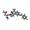  Mass: 498.522 Da / Num. of mol.: 3 / Source method: obtained synthetically / Formula: C26H28F2N4O4 / Feature type: SUBJECT OF INVESTIGATION Mass: 498.522 Da / Num. of mol.: 3 / Source method: obtained synthetically / Formula: C26H28F2N4O4 / Feature type: SUBJECT OF INVESTIGATION#4: Chemical | ChemComp-EDO / Ethylene glycol  Mass: 62.068 Da / Num. of mol.: 5 / Source method: obtained synthetically / Formula: C2H6O2 Mass: 62.068 Da / Num. of mol.: 5 / Source method: obtained synthetically / Formula: C2H6O2#5: Water | ChemComp-HOH / | Water Mass: 18.015 Da / Num. of mol.: 631 / Source method: isolated from a natural source / Formula: H2O Mass: 18.015 Da / Num. of mol.: 631 / Source method: isolated from a natural source / Formula: H2OHas ligand of interest | Y | |
|---|
-Experimental details
-Experiment
| Experiment | Method: X-RAY DIFFRACTION / Number of used crystals: 1 |
|---|
- Sample preparation
| Crystal | Density Matthews: 2.03 Å3/Da / Density % sol: 39.45 % |
|---|---|
| Crystal grow | Temperature: 293 K / Method: vapor diffusion, sitting drop / pH: 9 Details: 23.4mg/mL deglycosylated protein in 25mM HEPES/NaOH pH7, 300 mM NaCl, 200mM Trehalose incubated with 10-fold excess of ligand, then mixed 50-70% with 50-30% reservoir consisting of 25% v/v ...Details: 23.4mg/mL deglycosylated protein in 25mM HEPES/NaOH pH7, 300 mM NaCl, 200mM Trehalose incubated with 10-fold excess of ligand, then mixed 50-70% with 50-30% reservoir consisting of 25% v/v PEG smear broad, 0.1M Bicine/NaOH pH 9.0, 10% v/v 2-Propanol, total volume 200nL |
-Data collection
| Diffraction | Mean temperature: 100 K | ||||||||||||||||||||||||||||||||||||||||||||||||||||||||||||||||||||||||||||||||||||||||||||||||||||||||||||||||||||||||||||||||||||||||||||||||||||||||||||||||||||||||||||||||||||||||||||||||||||||||||||||||||
|---|---|---|---|---|---|---|---|---|---|---|---|---|---|---|---|---|---|---|---|---|---|---|---|---|---|---|---|---|---|---|---|---|---|---|---|---|---|---|---|---|---|---|---|---|---|---|---|---|---|---|---|---|---|---|---|---|---|---|---|---|---|---|---|---|---|---|---|---|---|---|---|---|---|---|---|---|---|---|---|---|---|---|---|---|---|---|---|---|---|---|---|---|---|---|---|---|---|---|---|---|---|---|---|---|---|---|---|---|---|---|---|---|---|---|---|---|---|---|---|---|---|---|---|---|---|---|---|---|---|---|---|---|---|---|---|---|---|---|---|---|---|---|---|---|---|---|---|---|---|---|---|---|---|---|---|---|---|---|---|---|---|---|---|---|---|---|---|---|---|---|---|---|---|---|---|---|---|---|---|---|---|---|---|---|---|---|---|---|---|---|---|---|---|---|---|---|---|---|---|---|---|---|---|---|---|---|---|---|---|---|---|
| Diffraction source | Source: SYNCHROTRON / Site: SLS / Beamline: X10SA / Wavelength: 0.99982 Å | ||||||||||||||||||||||||||||||||||||||||||||||||||||||||||||||||||||||||||||||||||||||||||||||||||||||||||||||||||||||||||||||||||||||||||||||||||||||||||||||||||||||||||||||||||||||||||||||||||||||||||||||||||
| Detector | Type: MARMOSAIC 225 mm CCD / Detector: CCD / Date: Mar 22, 2021 | ||||||||||||||||||||||||||||||||||||||||||||||||||||||||||||||||||||||||||||||||||||||||||||||||||||||||||||||||||||||||||||||||||||||||||||||||||||||||||||||||||||||||||||||||||||||||||||||||||||||||||||||||||
| Radiation | Protocol: SINGLE WAVELENGTH / Monochromatic (M) / Laue (L): M / Scattering type: x-ray | ||||||||||||||||||||||||||||||||||||||||||||||||||||||||||||||||||||||||||||||||||||||||||||||||||||||||||||||||||||||||||||||||||||||||||||||||||||||||||||||||||||||||||||||||||||||||||||||||||||||||||||||||||
| Radiation wavelength | Wavelength: 0.99982 Å / Relative weight: 1 | ||||||||||||||||||||||||||||||||||||||||||||||||||||||||||||||||||||||||||||||||||||||||||||||||||||||||||||||||||||||||||||||||||||||||||||||||||||||||||||||||||||||||||||||||||||||||||||||||||||||||||||||||||
| Reflection | Resolution: 1.45→70.37 Å / Num. obs: 207573 / % possible obs: 99.3 % / Redundancy: 3.447 % / Biso Wilson estimate: 31.763 Å2 / CC1/2: 0.999 / Rmerge(I) obs: 0.052 / Rrim(I) all: 0.062 / Χ2: 0.861 / Net I/σ(I): 9.71 / Num. measured all: 715549 / Scaling rejects: 143 | ||||||||||||||||||||||||||||||||||||||||||||||||||||||||||||||||||||||||||||||||||||||||||||||||||||||||||||||||||||||||||||||||||||||||||||||||||||||||||||||||||||||||||||||||||||||||||||||||||||||||||||||||||
| Reflection shell | Diffraction-ID: 1
|
- Processing
| Software |
| |||||||||||||||||||||||||||||||||||||||||||||||||||||||||||||||||||||||||||
|---|---|---|---|---|---|---|---|---|---|---|---|---|---|---|---|---|---|---|---|---|---|---|---|---|---|---|---|---|---|---|---|---|---|---|---|---|---|---|---|---|---|---|---|---|---|---|---|---|---|---|---|---|---|---|---|---|---|---|---|---|---|---|---|---|---|---|---|---|---|---|---|---|---|---|---|---|
| Refinement | Method to determine structure: MOLECULAR REPLACEMENT Starting model: inhouse model Resolution: 1.45→70.37 Å / Cor.coef. Fo:Fc: 0.968 / Cor.coef. Fo:Fc free: 0.958 / SU B: 1.36 / SU ML: 0.049 / Cross valid method: THROUGHOUT / σ(F): 0 / ESU R: 0.074 / ESU R Free: 0.074 / Stereochemistry target values: MAXIMUM LIKELIHOOD Details: Ligand in chain B has alternate conformations for the difluorophenyl
| |||||||||||||||||||||||||||||||||||||||||||||||||||||||||||||||||||||||||||
| Solvent computation | Ion probe radii: 0.8 Å / Shrinkage radii: 0.8 Å / VDW probe radii: 1.2 Å / Solvent model: MASK | |||||||||||||||||||||||||||||||||||||||||||||||||||||||||||||||||||||||||||
| Displacement parameters | Biso max: 80.63 Å2 / Biso mean: 21.328 Å2 / Biso min: 11.43 Å2
| |||||||||||||||||||||||||||||||||||||||||||||||||||||||||||||||||||||||||||
| Refinement step | Cycle: final / Resolution: 1.45→70.37 Å
| |||||||||||||||||||||||||||||||||||||||||||||||||||||||||||||||||||||||||||
| Refine LS restraints |
| |||||||||||||||||||||||||||||||||||||||||||||||||||||||||||||||||||||||||||
| LS refinement shell | Resolution: 1.445→1.483 Å / Rfactor Rfree error: 0 / Total num. of bins used: 20
|
