 Movie
Movie Controller

[English] 日本語
 Yorodumi
- PDB-7f3w: Crystal structure of cytochrome P450DA mutant (N190F/V356L/A486E)... -
+ Open data

- Basic information
| Entry | Database: PDB / ID: 7f3w | ||||||
|---|---|---|---|---|---|---|---|
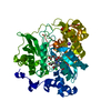
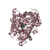
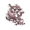
| Title | Crystal structure of cytochrome P450DA mutant (N190F/V356L/A486E) heme domain | ||||||
 Components Components | cytochrome P450 | ||||||
 Keywords Keywords | OXIDOREDUCTASE / cytochrome P450 | ||||||
| Function / homology | PROTOPORPHYRIN IX CONTAINING FE Function and homology information Function and homology information | ||||||
| Biological species |  Deinococcus apachensis (bacteria) Deinococcus apachensis (bacteria) | ||||||
| Method | X-RAY DIFFRACTION / SYNCHROTRON / MOLECULAR REPLACEMENT / Resolution: 2.86 Å | ||||||
 Authors Authors | Wan, N.W. | ||||||
 Citation Citation | Journal: To Be Published Title: Crystal structure of cytochrome P450DA mutant (N190F/V356L/A486E) heme domain Authors: Wan, N.W. | ||||||
| History |
|
- Structure visualization
| Structure viewer | Molecule: MolmilJmol/JSmol |
|---|
- Downloads & links
-Download
| PDBx/mmCIF format | 7f3w.cif.gz | 403.9 KB | Display | PDBx/mmCIF format |
|---|---|---|---|---|
| PDB format | pdb7f3w.ent.gz | 304.5 KB | Display | PDB format |
| PDBx/mmJSON format | 7f3w.json.gz | Tree view | PDBx/mmJSON format | |
| Others |  Other downloads Other downloads |
-Validation report
| Arichive directory | https://data.pdbj.org/pub/pdb/validation_reports/f3/7f3wftp://data.pdbj.org/pub/pdb/validation_reports/f3/7f3w | HTTPS FTP |
|---|
-Related structure data
| Related structure data |  1zoaS S: Starting model for refinement |
|---|---|
| Similar structure data |


-Links
 PDBj
PDBj
- Assembly
| Deposited unit | 
| ||||||||||||
|---|---|---|---|---|---|---|---|---|---|---|---|---|---|
| 1 | 
| ||||||||||||
| 2 | 
| ||||||||||||
| 3 | 
| ||||||||||||
| 4 | 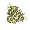
| ||||||||||||
| Unit cell |
|
-Components
| #1: Protein | Mass: 55969.648 Da / Num. of mol.: 4 Source method: isolated from a genetically manipulated source Source: (gene. exp.) Deinococcus apachensis (bacteria) / Production host: Escherichia coli (E. coli)#2: Chemical | ChemComp-HEM / Heme B  Mass: 616.487 Da / Num. of mol.: 4 / Source method: obtained synthetically / Formula: C34H32FeN4O4 Mass: 616.487 Da / Num. of mol.: 4 / Source method: obtained synthetically / Formula: C34H32FeN4O4#3: Chemical | ChemComp-SO4 / Sulfate  Mass: 96.063 Da / Num. of mol.: 10 / Source method: obtained synthetically / Formula: SO4 Mass: 96.063 Da / Num. of mol.: 10 / Source method: obtained synthetically / Formula: SO4Has ligand of interest | N | |
|---|
-Experimental details
-Experiment
| Experiment | Method: X-RAY DIFFRACTION / Number of used crystals: 1 |
|---|
- Sample preparation
| Crystal | Density Matthews: 2.92 Å3/Da / Density % sol: 57.91 % |
|---|---|
| Crystal grow | Temperature: 293 K / Method: vapor diffusion, hanging drop / Details: 0.1 M MES 6.0, 10 % w/v Polyethylene glycol 4000 |
-Data collection
| Diffraction | Mean temperature: 100 K / Serial crystal experiment: N |
|---|---|
| Diffraction source | Source: SYNCHROTRON / Site: SSRF  / Beamline: BL18U1 / Wavelength: 0.9791 Å / Beamline: BL18U1 / Wavelength: 0.9791 Å |
| Detector | Type: DECTRIS PILATUS 6M / Detector: PIXEL / Date: Oct 27, 2020 |
| Radiation | Protocol: SINGLE WAVELENGTH / Monochromatic (M) / Laue (L): M / Scattering type: x-ray |
| Radiation wavelength | Wavelength: 0.9791 Å / Relative weight: 1 |
| Reflection | Resolution: 2.86→50 Å / Num. obs: 80451 / % possible obs: 91.9 % / Redundancy: 9.6 % / Biso Wilson estimate: 45.62 Å2 / Rmerge(I) obs: 0.161 / Rpim(I) all: 0.068 / Net I/σ(I): 11 |
| Reflection shell | Resolution: 2.86→2.92 Å / Rmerge(I) obs: 0.751 / Mean I/σ(I) obs: 2.2 / Num. unique obs: 2460 / Rpim(I) all: 0.342 |
- Processing
| Software |
| |||||||||||||||||||||||||||||||||||||||||||||||||||||||||||||||||||||||||||||||||||||||||||||||||||||||||||||||||||||||||||||||||||||||||||||||||||||||||||||||||
|---|---|---|---|---|---|---|---|---|---|---|---|---|---|---|---|---|---|---|---|---|---|---|---|---|---|---|---|---|---|---|---|---|---|---|---|---|---|---|---|---|---|---|---|---|---|---|---|---|---|---|---|---|---|---|---|---|---|---|---|---|---|---|---|---|---|---|---|---|---|---|---|---|---|---|---|---|---|---|---|---|---|---|---|---|---|---|---|---|---|---|---|---|---|---|---|---|---|---|---|---|---|---|---|---|---|---|---|---|---|---|---|---|---|---|---|---|---|---|---|---|---|---|---|---|---|---|---|---|---|---|---|---|---|---|---|---|---|---|---|---|---|---|---|---|---|---|---|---|---|---|---|---|---|---|---|---|---|---|---|---|---|---|
| Refinement | Method to determine structure: MOLECULAR REPLACEMENT Starting model: 1zoa Resolution: 2.86→29.14 Å / SU ML: 0.3348 / Cross valid method: FREE R-VALUE / σ(F): 1.34 / Phase error: 24.5234 Stereochemistry target values: GeoStd + Monomer Library + CDL v1.2
| |||||||||||||||||||||||||||||||||||||||||||||||||||||||||||||||||||||||||||||||||||||||||||||||||||||||||||||||||||||||||||||||||||||||||||||||||||||||||||||||||
| Solvent computation | Shrinkage radii: 0.9 Å / VDW probe radii: 1.11 Å / Solvent model: FLAT BULK SOLVENT MODEL | |||||||||||||||||||||||||||||||||||||||||||||||||||||||||||||||||||||||||||||||||||||||||||||||||||||||||||||||||||||||||||||||||||||||||||||||||||||||||||||||||
| Displacement parameters | Biso mean: 45.1 Å2 | |||||||||||||||||||||||||||||||||||||||||||||||||||||||||||||||||||||||||||||||||||||||||||||||||||||||||||||||||||||||||||||||||||||||||||||||||||||||||||||||||
| Refinement step | Cycle: LAST / Resolution: 2.86→29.14 Å
| |||||||||||||||||||||||||||||||||||||||||||||||||||||||||||||||||||||||||||||||||||||||||||||||||||||||||||||||||||||||||||||||||||||||||||||||||||||||||||||||||
| Refine LS restraints |
| |||||||||||||||||||||||||||||||||||||||||||||||||||||||||||||||||||||||||||||||||||||||||||||||||||||||||||||||||||||||||||||||||||||||||||||||||||||||||||||||||
| LS refinement shell |
|
