 Movie
Movie Controller

[English] 日本語
 Yorodumi
- PDB-7ezw: Cyclic Peptide that Interacts with the eIF4E Capped-mRNA Binding Site -
+ Open data

- Basic information
| Entry | Database: PDB / ID: 7ezw | ||||||
|---|---|---|---|---|---|---|---|
| Title | Cyclic Peptide that Interacts with the eIF4E Capped-mRNA Binding Site | ||||||
 Components Components |
| ||||||
 Keywords Keywords | RNA BINDING PROTEIN/INHIBITOR / ap dependent translation / RNA BINDING PROTEIN-INHIBITOR COMPLEX | ||||||
| Function / homology |  Function and homology information Function and homology informationActivation of the mRNA upon binding of the cap-binding complex and eIFs, and subsequent binding to 43S / : /  eukaryotic initiation factor 4G binding / regulation of translation at postsynapse, modulating synaptic transmission / RNA cap binding / chromatoid body / eukaryotic translation initiation factor 4F complex / Z-decay: degradation of maternal mRNAs by zygotically expressed factors / mRNA cap binding / Deadenylation of mRNA ...Activation of the mRNA upon binding of the cap-binding complex and eIFs, and subsequent binding to 43S / : / eukaryotic initiation factor 4G binding / regulation of translation at postsynapse, modulating synaptic transmission / RNA cap binding / chromatoid body / eukaryotic translation initiation factor 4F complex / Z-decay: degradation of maternal mRNAs by zygotically expressed factors / mRNA cap binding / Deadenylation of mRNA / Transport of the SLBP independent Mature mRNA / RNA 7-methylguanosine cap binding / Transport of the SLBP Dependant Mature mRNA / M-decay: degradation of maternal mRNAs by maternally stored factors / Transport of Mature mRNA Derived from an Intronless Transcript / RISC complex / Ribosomal scanning and start codon recognition / Translation initiation complex formation / stem cell population maintenance / mTORC1-mediated signalling / GTP hydrolysis and joining of the 60S ribosomal subunit / negative regulation of neuron differentiation / L13a-mediated translational silencing of Ceruloplasmin expression / behavioral fear response / mRNA export from nucleus / translational initiation / translation initiation factor activity / positive regulation of mitotic cell cycle / cellular response to dexamethasone stimulus / P-body / neuron differentiation / G1/S transition of mitotic cell cycle / ISG15 antiviral mechanism / cytoplasmic stress granule / cytoplasmic ribonucleoprotein granule / regulation of translation / postsynapse / DNA-binding transcription factor binding / negative regulation of translation / nuclear speck / glutamatergic synapse / perinuclear region of cytoplasm / enzyme binding / RNA binding / extracellular exosome / nucleus / cytosol / cytoplasm eukaryotic initiation factor 4G binding / regulation of translation at postsynapse, modulating synaptic transmission / RNA cap binding / chromatoid body / eukaryotic translation initiation factor 4F complex / Z-decay: degradation of maternal mRNAs by zygotically expressed factors / mRNA cap binding / Deadenylation of mRNA ...Activation of the mRNA upon binding of the cap-binding complex and eIFs, and subsequent binding to 43S / : / eukaryotic initiation factor 4G binding / regulation of translation at postsynapse, modulating synaptic transmission / RNA cap binding / chromatoid body / eukaryotic translation initiation factor 4F complex / Z-decay: degradation of maternal mRNAs by zygotically expressed factors / mRNA cap binding / Deadenylation of mRNA / Transport of the SLBP independent Mature mRNA / RNA 7-methylguanosine cap binding / Transport of the SLBP Dependant Mature mRNA / M-decay: degradation of maternal mRNAs by maternally stored factors / Transport of Mature mRNA Derived from an Intronless Transcript / RISC complex / Ribosomal scanning and start codon recognition / Translation initiation complex formation / stem cell population maintenance / mTORC1-mediated signalling / GTP hydrolysis and joining of the 60S ribosomal subunit / negative regulation of neuron differentiation / L13a-mediated translational silencing of Ceruloplasmin expression / behavioral fear response / mRNA export from nucleus / translational initiation / translation initiation factor activity / positive regulation of mitotic cell cycle / cellular response to dexamethasone stimulus / P-body / neuron differentiation / G1/S transition of mitotic cell cycle / ISG15 antiviral mechanism / cytoplasmic stress granule / cytoplasmic ribonucleoprotein granule / regulation of translation / postsynapse / DNA-binding transcription factor binding / negative regulation of translation / nuclear speck / glutamatergic synapse / perinuclear region of cytoplasm / enzyme binding / RNA binding / extracellular exosome / nucleus / cytosol / cytoplasmSimilarity search - Function | ||||||
| Biological species |  Homo sapiens (human) Homo sapiens (human)synthetic construct (others) | ||||||
| Method | X-RAY DIFFRACTION / SYNCHROTRON / MOLECULAR REPLACEMENT / Resolution: 2.35 Å | ||||||
 Authors Authors | Brown, C.J. / Ng, S. / Frosi, Y. | ||||||
 Citation Citation | Journal: Rsc Chem Biol / Year: 2022 Title: Development of a novel peptide aptamer that interacts with the eIF4E capped-mRNA binding site using peptide epitope linker evolution (PELE). Authors: Frosi, Y. / Ng, S. / Lin, Y.C. / Jiang, S. / Ramlan, S.R. / Lama, D. / Verma, C.S. / Asial, I. / Brown, C.J. | ||||||
| History |
|
- Structure visualization
| Structure viewer | Molecule: MolmilJmol/JSmol |
|---|
- Downloads & links
-Download
| PDBx/mmCIF format | 7ezw.cif.gz | 54.6 KB | Display | PDBx/mmCIF format |
|---|---|---|---|---|
| PDB format | pdb7ezw.ent.gz | 37.3 KB | Display | PDB format |
| PDBx/mmJSON format | 7ezw.json.gz | Tree view | PDBx/mmJSON format | |
| Others |  Other downloads Other downloads |
-Validation report
| Arichive directory | https://data.pdbj.org/pub/pdb/validation_reports/ez/7ezwftp://data.pdbj.org/pub/pdb/validation_reports/ez/7ezw | HTTPS FTP |
|---|
-Related structure data
| Related structure data | 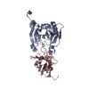 7f07C 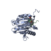 4beaS S: Starting model for refinement C: citing same article ( |
|---|---|
| Similar structure data |
-Links
 PDBj
PDBj


- Assembly
| Deposited unit | 
| ||||||||
|---|---|---|---|---|---|---|---|---|---|
| 1 |
| ||||||||
| Unit cell |
| ||||||||
| Components on special symmetry positions |
|
-Components
| #1: Protein | EIF4E / eIF4E / eIF-4F 25 kDa subunit / mRNA cap-binding protein Mass: 25130.242 Da / Num. of mol.: 1 Source method: isolated from a genetically manipulated source Source: (gene. exp.) Homo sapiens (human) / Gene: EIF4E, EIF4EL1, EIF4F / Production host:  Escherichia coli (E. coli) / References: UniProt: P06730 Escherichia coli (E. coli) / References: UniProt: P06730 |
|---|---|
| #2: Protein/peptide | Mass: 1205.364 Da / Num. of mol.: 1 / Source method: obtained synthetically / Details: amidation / Source: (synth.) synthetic construct (others) |
| #3: Chemical | ChemComp-NA /   Mass: 22.990 Da / Num. of mol.: 1 / Source method: obtained synthetically / Formula: Na Mass: 22.990 Da / Num. of mol.: 1 / Source method: obtained synthetically / Formula: Na |
| #4: Water | ChemComp-HOH / Water Mass: 18.015 Da / Num. of mol.: 59 / Source method: obtained synthetically / Formula: H2O Mass: 18.015 Da / Num. of mol.: 59 / Source method: obtained synthetically / Formula: H2O |
| Has ligand of interest | Y |
-Experimental details
-Experiment
| Experiment | Method: X-RAY DIFFRACTION / Number of used crystals: 1 |
|---|
- Sample preparation
| Crystal | Density Matthews: 2.39 Å3/Da / Density % sol: 48.43 % |
|---|---|
| Crystal grow | Temperature: 293 K / Method: vapor diffusion, sitting drop / Details: 0.2 M Potassium chloride 20% (w/v) PEG 3350 |
-Data collection
| Diffraction | Mean temperature: 100 K / Serial crystal experiment: N |
|---|---|
| Diffraction source | Source: SYNCHROTRON / Site: Australian Synchrotron  / Beamline: MX1 / Wavelength: 0.9537 Å / Beamline: MX1 / Wavelength: 0.9537 Å |
| Detector | Type: DECTRIS EIGER X 9M / Detector: PIXEL / Date: Mar 29, 2017 |
| Radiation | Protocol: SINGLE WAVELENGTH / Monochromatic (M) / Laue (L): M / Scattering type: x-ray |
| Radiation wavelength | Wavelength: 0.9537 Å / Relative weight: 1 |
| Reflection | Resolution: 2.35→48.194 Å / Num. obs: 10511 / % possible obs: 99.9 % / Redundancy: 11.3 % / Rmerge(I) obs: 0.114 / Net I/σ(I): 13.8 |
| Reflection shell | Resolution: 2.35→2.47 Å / Redundancy: 11.2 % / Rmerge(I) obs: 0.711 / Mean I/σ(I) obs: 2.6 / % possible all: 99.2 |
- Processing
| Software |
| ||||||||||||||||||||||||||||||||||||||||||||||||||||||||||||||||||||||||||||||||||||||||||||||||||||||||||||||||||||||||||||||||||||||||||||||||||||||||||||||||||||||||||||||||||||||
|---|---|---|---|---|---|---|---|---|---|---|---|---|---|---|---|---|---|---|---|---|---|---|---|---|---|---|---|---|---|---|---|---|---|---|---|---|---|---|---|---|---|---|---|---|---|---|---|---|---|---|---|---|---|---|---|---|---|---|---|---|---|---|---|---|---|---|---|---|---|---|---|---|---|---|---|---|---|---|---|---|---|---|---|---|---|---|---|---|---|---|---|---|---|---|---|---|---|---|---|---|---|---|---|---|---|---|---|---|---|---|---|---|---|---|---|---|---|---|---|---|---|---|---|---|---|---|---|---|---|---|---|---|---|---|---|---|---|---|---|---|---|---|---|---|---|---|---|---|---|---|---|---|---|---|---|---|---|---|---|---|---|---|---|---|---|---|---|---|---|---|---|---|---|---|---|---|---|---|---|---|---|---|---|
| Refinement | Method to determine structure: MOLECULAR REPLACEMENT Starting model: 4BEA Resolution: 2.35→48.19 Å / Cor.coef. Fo:Fc: 0.936 / Cor.coef. Fo:Fc free: 0.91 / SU B: 7.698 / SU ML: 0.178 / Cross valid method: FREE R-VALUE / ESU R: 0.332 / ESU R Free: 0.238 Details: HYDROGENS HAVE BEEN ADDED IN THEIR RIDING POSITIONS
| ||||||||||||||||||||||||||||||||||||||||||||||||||||||||||||||||||||||||||||||||||||||||||||||||||||||||||||||||||||||||||||||||||||||||||||||||||||||||||||||||||||||||||||||||||||||
| Solvent computation | Ion probe radii: 0.8 Å / Shrinkage radii: 0.8 Å / VDW probe radii: 1.2 Å / Solvent model: MASK BULK SOLVENT | ||||||||||||||||||||||||||||||||||||||||||||||||||||||||||||||||||||||||||||||||||||||||||||||||||||||||||||||||||||||||||||||||||||||||||||||||||||||||||||||||||||||||||||||||||||||
| Displacement parameters | Biso mean: 33.22 Å2
| ||||||||||||||||||||||||||||||||||||||||||||||||||||||||||||||||||||||||||||||||||||||||||||||||||||||||||||||||||||||||||||||||||||||||||||||||||||||||||||||||||||||||||||||||||||||
| Refinement step | Cycle: LAST / Resolution: 2.35→48.19 Å
| ||||||||||||||||||||||||||||||||||||||||||||||||||||||||||||||||||||||||||||||||||||||||||||||||||||||||||||||||||||||||||||||||||||||||||||||||||||||||||||||||||||||||||||||||||||||
| Refine LS restraints |
|