 Movie
Movie Controller


+ Open data

- Basic information
| Entry | Database: PDB / ID: 7exb | ||||||
|---|---|---|---|---|---|---|---|
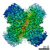
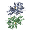
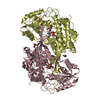
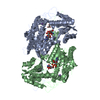
| Title | DfgA-DfgB complex apo 2.4 angstrom | ||||||
 Components Components |
| ||||||
 Keywords Keywords |  LYASE / C-deglycosylation / sugar phosphate isomerase/epimerase LYASE / C-deglycosylation / sugar phosphate isomerase/epimerase | ||||||
| Function / homology | Domain of unknown function DUF6379 / Domain of unknown function (DUF6379) / Xylose isomerase-like, TIM barrel domain / Xylose isomerase-like TIM barrel / Xylose isomerase-like superfamily / isomerase activity / : / DUF6379 domain-containing protein / Sugar phosphate isomerase/epimerase Function and homology information Function and homology information | ||||||
| Biological species |  [Eubacterium] cellulosolvens 6 (bacteria) [Eubacterium] cellulosolvens 6 (bacteria) | ||||||
| Method | X-RAY DIFFRACTION / SYNCHROTRON / MOLECULAR REPLACEMENT / Resolution: 2.4 Å | ||||||
 Authors Authors | Mori, T. / Senda, M. / Senda, T. / Abe, I. | ||||||
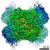
 Citation Citation | Journal: Nat Commun / Year: 2021 Title: C-Glycoside metabolism in the gut and in nature: Identification, characterization, structural analyses and distribution of C-C bond-cleaving enzymes. Authors: Takahiro Mori / Takuto Kumano / Haibing He / Satomi Watanabe / Miki Senda / Toshio Moriya / Naruhiko Adachi / Sanae Hori / Yuzu Terashita / Masato Kawasaki / Yoshiteru Hashimoto / Takayoshi ...Authors: Takahiro Mori / Takuto Kumano / Haibing He / Satomi Watanabe / Miki Senda / Toshio Moriya / Naruhiko Adachi / Sanae Hori / Yuzu Terashita / Masato Kawasaki / Yoshiteru Hashimoto / Takayoshi Awakawa / Toshiya Senda / Ikuro Abe / Michihiko Kobayashi /  Abstract: C-Glycosides, in which a sugar moiety is linked via a carbon-carbon (C-C) bond to a non-sugar moiety (aglycone), are found in our food and medicine. The C-C bond is cleaved by intestinal microbes and ...C-Glycosides, in which a sugar moiety is linked via a carbon-carbon (C-C) bond to a non-sugar moiety (aglycone), are found in our food and medicine. The C-C bond is cleaved by intestinal microbes and the resulting aglycones exert various bioactivities. Although the enzymes responsible for the reactions have been identified, their catalytic mechanisms and the generality of the reactions in nature remain to be explored. Here, we present the identification and structural basis for the activation of xenobiotic C-glycosides by heterocomplex C-deglycosylation enzymes from intestinal and soil bacteria. They are found to be metal-dependent enzymes exhibiting broad substrate specificity toward C-glycosides. X-ray crystallographic and cryo-electron microscopic analyses, as well as structure-based mutagenesis, reveal the structural details of these enzymes and the detailed catalytic mechanisms of their remarkable C-C bond cleavage reactions. Furthermore, bioinformatic and biochemical analyses suggest that the C-deglycosylation enzymes are widely distributed in the gut, soil, and marine bacteria. | ||||||
| History |
|
- Structure visualization
| Structure viewer | Molecule: MolmilJmol/JSmol |
|---|
- Downloads & links
-Download
| PDBx/mmCIF format | 7exb.cif.gz | 136.3 KB | Display | PDBx/mmCIF format |
|---|---|---|---|---|
| PDB format | pdb7exb.ent.gz | 84 KB | Display | PDB format |
| PDBx/mmJSON format | 7exb.json.gz | Tree view | PDBx/mmJSON format | |
| Others |  Other downloads Other downloads |
-Validation report
| Arichive directory | https://data.pdbj.org/pub/pdb/validation_reports/ex/7exbftp://data.pdbj.org/pub/pdb/validation_reports/ex/7exb | HTTPS FTP |
|---|
-Related structure data
| Related structure data |  7bvrC 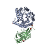 7bvsSC 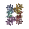 7drdC 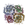 7dreC  7exzC S: Starting model for refinement C: citing same article ( |
|---|---|
| Similar structure data |


-Links
 PDBj
PDBj
- Assembly
| Deposited unit | 
| ||||||||||||
|---|---|---|---|---|---|---|---|---|---|---|---|---|---|
| 1 | 
| ||||||||||||
| Unit cell |
|
-Components
| #1: Protein | Mass: 33764.246 Da / Num. of mol.: 1 Source method: isolated from a genetically manipulated source Source: (gene. exp.) [Eubacterium] cellulosolvens 6 (bacteria)Gene: EubceDRAFT1_2664 / Production host: Escherichia coli (E. coli) / References: UniProt: I5AX50 | ||||
|---|---|---|---|---|---|
| #2: Protein | Mass: 18458.150 Da / Num. of mol.: 1 Source method: isolated from a genetically manipulated source Source: (gene. exp.) [Eubacterium] cellulosolvens 6 (bacteria)Gene: EubceDRAFT1_2663 / Production host: Escherichia coli (E. coli) / References: UniProt: I5AX49 | ||||
| #3: Chemical | ChemComp-MN /   Mass: 54.938 Da / Num. of mol.: 1 / Source method: obtained synthetically / Formula: Mn / Feature type: SUBJECT OF INVESTIGATION Mass: 54.938 Da / Num. of mol.: 1 / Source method: obtained synthetically / Formula: Mn / Feature type: SUBJECT OF INVESTIGATION | ||||
| #4: Chemical | ChemComp-SO4 / Sulfate  Mass: 96.063 Da / Num. of mol.: 4 / Source method: obtained synthetically / Formula: SO4 Mass: 96.063 Da / Num. of mol.: 4 / Source method: obtained synthetically / Formula: SO4#5: Water | ChemComp-HOH / | Water Mass: 18.015 Da / Num. of mol.: 329 / Source method: isolated from a natural source / Formula: H2O Mass: 18.015 Da / Num. of mol.: 329 / Source method: isolated from a natural source / Formula: H2OHas ligand of interest | Y | |
-Experimental details
-Experiment
| Experiment | Method: X-RAY DIFFRACTION / Number of used crystals: 1 |
|---|
- Sample preparation
| Crystal | Density Matthews: 6.06 Å3/Da / Density % sol: 79.7 % |
|---|---|
| Crystal grow | Temperature: 293 K / Method: vapor diffusion, sitting drop / Details: 100 mM Tris-HCl (pH8.5), 1190 mM (NH4)2SO4 |
-Data collection
| Diffraction | Mean temperature: 100 K / Serial crystal experiment: N |
|---|---|
| Diffraction source | Source: SYNCHROTRON / Site: Photon Factory / Beamline: BL-17A / Wavelength: 1.88 Å |
| Detector | Type: DECTRIS EIGER X 16M / Detector: PIXEL / Date: May 19, 2021 |
| Radiation | Protocol: SINGLE WAVELENGTH / Monochromatic (M) / Laue (L): M / Scattering type: x-ray |
| Radiation wavelength | Wavelength: 1.88 Å / Relative weight: 1 |
| Reflection | Resolution: 2.4→47.1 Å / Num. obs: 51290 / % possible obs: 100 % / Redundancy: 36.9 % / Biso Wilson estimate: 43.75 Å2 / CC1/2: 1 / Rmerge(I) obs: 0.14 / Net I/σ(I): 27.4 |
| Reflection shell | Resolution: 2.4→2.47 Å / Redundancy: 22.8 % / Rmerge(I) obs: 1.417 / Mean I/σ(I) obs: 2.8 / Num. unique obs: 4359 / CC1/2: 0.83 / % possible all: 99.9 |
- Processing
| Software |
| |||||||||||||||||||||||||||||||||||||||||||||||||||||||||||||||||||||||||||||||||||||||||||||||||||||||||||||||||||||||||||||||||||||
|---|---|---|---|---|---|---|---|---|---|---|---|---|---|---|---|---|---|---|---|---|---|---|---|---|---|---|---|---|---|---|---|---|---|---|---|---|---|---|---|---|---|---|---|---|---|---|---|---|---|---|---|---|---|---|---|---|---|---|---|---|---|---|---|---|---|---|---|---|---|---|---|---|---|---|---|---|---|---|---|---|---|---|---|---|---|---|---|---|---|---|---|---|---|---|---|---|---|---|---|---|---|---|---|---|---|---|---|---|---|---|---|---|---|---|---|---|---|---|---|---|---|---|---|---|---|---|---|---|---|---|---|---|---|---|
| Refinement | Method to determine structure: MOLECULAR REPLACEMENT Starting model: 7BVS Resolution: 2.4→47.1 Å / SU ML: 0.2941 / Cross valid method: FREE R-VALUE / σ(F): 1.34 / Phase error: 20.4611 Stereochemistry target values: GeoStd + Monomer Library + CDL v1.2
| |||||||||||||||||||||||||||||||||||||||||||||||||||||||||||||||||||||||||||||||||||||||||||||||||||||||||||||||||||||||||||||||||||||
| Solvent computation | Shrinkage radii: 0.9 Å / VDW probe radii: 1.11 Å / Solvent model: FLAT BULK SOLVENT MODEL | |||||||||||||||||||||||||||||||||||||||||||||||||||||||||||||||||||||||||||||||||||||||||||||||||||||||||||||||||||||||||||||||||||||
| Displacement parameters | Biso mean: 42.02 Å2 | |||||||||||||||||||||||||||||||||||||||||||||||||||||||||||||||||||||||||||||||||||||||||||||||||||||||||||||||||||||||||||||||||||||
| Refinement step | Cycle: LAST / Resolution: 2.4→47.1 Å
| |||||||||||||||||||||||||||||||||||||||||||||||||||||||||||||||||||||||||||||||||||||||||||||||||||||||||||||||||||||||||||||||||||||
| Refine LS restraints |
| |||||||||||||||||||||||||||||||||||||||||||||||||||||||||||||||||||||||||||||||||||||||||||||||||||||||||||||||||||||||||||||||||||||
| LS refinement shell |
|
