 Movie
Movie Controller


+ Open data

- Basic information
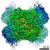
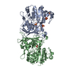
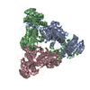
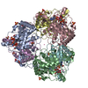
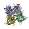
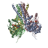
| Entry | Database: PDB / ID: 7dre | ||||||
|---|---|---|---|---|---|---|---|
| Title | Cryo-EM structure of DfgA-B at 2.54 angstrom resolution | ||||||
 Components Components |
| ||||||
 Keywords Keywords |  BIOSYNTHETIC PROTEIN / C-deglycosylase / sugar-isomerase-like BIOSYNTHETIC PROTEIN / C-deglycosylase / sugar-isomerase-like | ||||||
| Function / homology | Domain of unknown function DUF6379 / Domain of unknown function (DUF6379) / Xylose isomerase-like, TIM barrel domain / Xylose isomerase-like TIM barrel / Xylose isomerase-like superfamily / isomerase activity / DUF6379 domain-containing protein / Sugar phosphate isomerase/epimerase Function and homology information Function and homology information | ||||||
| Biological species |  [Eubacterium] cellulosolvens 6 (bacteria) [Eubacterium] cellulosolvens 6 (bacteria) | ||||||
| Method | ELECTRON MICROSCOPY / single particle reconstruction / cryo EM / Resolution: 2.54 Å | ||||||
 Authors Authors | Mori, T. / Moriya, T. / Adachi, N. / Senda, T. / Abe, I. | ||||||
| Funding support |  Japan, 1items Japan, 1items
| ||||||
 Citation Citation | Journal: Nat Commun / Year: 2021 Title: C-Glycoside metabolism in the gut and in nature: Identification, characterization, structural analyses and distribution of C-C bond-cleaving enzymes. Authors: Takahiro Mori / Takuto Kumano / Haibing He / Satomi Watanabe / Miki Senda / Toshio Moriya / Naruhiko Adachi / Sanae Hori / Yuzu Terashita / Masato Kawasaki / Yoshiteru Hashimoto / Takayoshi ...Authors: Takahiro Mori / Takuto Kumano / Haibing He / Satomi Watanabe / Miki Senda / Toshio Moriya / Naruhiko Adachi / Sanae Hori / Yuzu Terashita / Masato Kawasaki / Yoshiteru Hashimoto / Takayoshi Awakawa / Toshiya Senda / Ikuro Abe / Michihiko Kobayashi / Abstract: C-Glycosides, in which a sugar moiety is linked via a carbon-carbon (C-C) bond to a non-sugar moiety (aglycone), are found in our food and medicine. The C-C bond is cleaved by intestinal microbes and ...C-Glycosides, in which a sugar moiety is linked via a carbon-carbon (C-C) bond to a non-sugar moiety (aglycone), are found in our food and medicine. The C-C bond is cleaved by intestinal microbes and the resulting aglycones exert various bioactivities. Although the enzymes responsible for the reactions have been identified, their catalytic mechanisms and the generality of the reactions in nature remain to be explored. Here, we present the identification and structural basis for the activation of xenobiotic C-glycosides by heterocomplex C-deglycosylation enzymes from intestinal and soil bacteria. They are found to be metal-dependent enzymes exhibiting broad substrate specificity toward C-glycosides. X-ray crystallographic and cryo-electron microscopic analyses, as well as structure-based mutagenesis, reveal the structural details of these enzymes and the detailed catalytic mechanisms of their remarkable C-C bond cleavage reactions. Furthermore, bioinformatic and biochemical analyses suggest that the C-deglycosylation enzymes are widely distributed in the gut, soil, and marine bacteria. | ||||||
| History |
|
- Structure visualization
| Movie |
Movie viewer |
|---|---|
| Structure viewer | Molecule: MolmilJmol/JSmol |
- Downloads & links
-Download
| PDBx/mmCIF format | 7dre.cif.gz | 273.7 KB | Display | PDBx/mmCIF format |
|---|---|---|---|---|
| PDB format | pdb7dre.ent.gz | 229.8 KB | Display | PDB format |
| PDBx/mmJSON format | 7dre.json.gz | Tree view | PDBx/mmJSON format | |
| Others |  Other downloads Other downloads |
-Validation report
| Arichive directory | https://data.pdbj.org/pub/pdb/validation_reports/dr/7dreftp://data.pdbj.org/pub/pdb/validation_reports/dr/7dre | HTTPS FTP |
|---|
-Related structure data
| Related structure data | 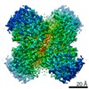 30809MC  7bvrC 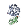 7bvsC 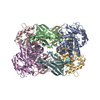 7drdC 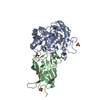 7exbC  7exzC M: map data used to model this data C: citing same article ( |
|---|---|
| Similar structure data |


-Links
 PDBj
PDBj
- Assembly
| Deposited unit | 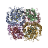
|
|---|---|
| 1 |
|
-Components
| #1: Protein | Mass: 33764.246 Da / Num. of mol.: 4 Source method: isolated from a genetically manipulated source Source: (gene. exp.) [Eubacterium] cellulosolvens 6 (bacteria)Gene: EubceDRAFT1_2664 / Production host: Escherichia coli (E. coli) / References: UniProt: I5AX50#2: Protein | Mass: 18458.150 Da / Num. of mol.: 4 Source method: isolated from a genetically manipulated source Source: (gene. exp.) [Eubacterium] cellulosolvens 6 (bacteria)Gene: EubceDRAFT1_2663 / Production host: Escherichia coli (E. coli) / References: UniProt: I5AX49 |
|---|
-Experimental details
-Experiment
| Experiment | Method: ELECTRON MICROSCOPY |
|---|---|
| EM experiment | Aggregation state: PARTICLE / 3D reconstruction method: single particle reconstruction |
- Sample preparation
| Component | Name: DfgA and DfgB / Type: COMPLEX / Details: heterodimer complex of DfgA and DfgB / Entity ID: all / Source: RECOMBINANT | |||||||||||||||
|---|---|---|---|---|---|---|---|---|---|---|---|---|---|---|---|---|
| Molecular weight | Value: 0.21 MDa / Experimental value: NO | |||||||||||||||
| Source (natural) | Organism: [Eubacterium] cellulosolvens (bacteria) | |||||||||||||||
| Source (recombinant) | Organism: Escherichia coli (E. coli) | |||||||||||||||
| Buffer solution | pH: 7.5 | |||||||||||||||
| Buffer component |
| |||||||||||||||
| Specimen | Conc.: 2 mg/ml / Embedding applied: NO / Shadowing applied: NO / Staining applied: NO / Vitrification applied: YES / Details: This sample was mono-disperse. | |||||||||||||||
| Specimen support | Details: The grid was washed by acetone prior to use. / Grid material: COPPER / Grid mesh size: 300 divisions/in. / Grid type: Quantifoil R1.2/1.3 | |||||||||||||||
| Vitrification | Instrument: FEI VITROBOT MARK IV / Cryogen name: ETHANE / Humidity: 100 % / Chamber temperature: 291 K / Details: Blotting time was 20 seconds (blot force 0) |
- Electron microscopy imaging
| Microscopy | Model: TFS TALOS |
|---|---|
| Electron gun | Electron source: FIELD EMISSION GUN / Accelerating voltage: 200 kV / Illumination mode: FLOOD BEAM |
| Electron lens | Mode: BRIGHT FIELDBright-field microscopy / Nominal magnification: 150000 X / Nominal defocus max: 1500 nm / Nominal defocus min: 600 nm / Cs: 2.7 mm / C2 aperture diameter: 50 µm |
| Specimen holder | Cryogen: NITROGEN |
| Image recording | Average exposure time: 62.06 sec. / Electron dose: 50 e/Å2 / Detector mode: COUNTING / Film or detector model: FEI FALCON III (4k x 4k) / Num. of grids imaged: 1 / Num. of real images: 1664 |
- Processing
| Software | Name: PHENIX / Version: 1.18.2_3874: / Classification: refinement | ||||||||||||||||||||||||||||||||||||||||||||||||||
|---|---|---|---|---|---|---|---|---|---|---|---|---|---|---|---|---|---|---|---|---|---|---|---|---|---|---|---|---|---|---|---|---|---|---|---|---|---|---|---|---|---|---|---|---|---|---|---|---|---|---|---|
| EM software |
| ||||||||||||||||||||||||||||||||||||||||||||||||||
| CTF correction | Type: PHASE FLIPPING AND AMPLITUDE CORRECTION | ||||||||||||||||||||||||||||||||||||||||||||||||||
| Particle selection | Num. of particles selected: 330866 | ||||||||||||||||||||||||||||||||||||||||||||||||||
| Symmetry | Point symmetry: D2 (2x2 fold dihedral) | ||||||||||||||||||||||||||||||||||||||||||||||||||
| 3D reconstruction | Resolution: 2.54 Å / Resolution method: FSC 0.143 CUT-OFF / Num. of particles: 60587 / Algorithm: FOURIER SPACE / Symmetry type: POINT | ||||||||||||||||||||||||||||||||||||||||||||||||||
| Atomic model building | Protocol: OTHER / Space: REAL | ||||||||||||||||||||||||||||||||||||||||||||||||||
| Refine LS restraints |
|
