 Movie
Movie Controller

[English] 日本語
 Yorodumi
- PDB-7ewo: Crystal Structure of D67A, E68P double mutant of O-acetyl-L-serin... -
+ Open data

- Basic information
| Entry | Database: PDB / ID: 7ewo | ||||||
|---|---|---|---|---|---|---|---|
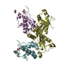
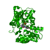
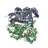
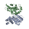
| Title | Crystal Structure of D67A, E68P double mutant of O-acetyl-L-serine sulfhydrylase from Haemophilus influenzae | ||||||
 Components Components | Cysteine synthase | ||||||
 Keywords Keywords | TRANSFERASE / Evolution / Cysteine Synthase / Protein-Protein interaction | ||||||
| Function / homology |  Function and homology informationcysteine synthase / cystathionine beta-synthase activity / cysteine synthase activity / L-cysteine desulfhydrase activity / cysteine biosynthetic process from serine / cytoplasm Function and homology informationcysteine synthase / cystathionine beta-synthase activity / cysteine synthase activity / L-cysteine desulfhydrase activity / cysteine biosynthetic process from serine / cytoplasmSimilarity search - Function | ||||||
| Biological species |  Haemophilus influenzae (bacteria) Haemophilus influenzae (bacteria) | ||||||
| Method | X-RAY DIFFRACTION / MOLECULAR REPLACEMENT / Resolution: 2.4 Å | ||||||
 Authors Authors | Rahisuddin, R. / Ekka, M.K. / Singh, A.K. / Saini, N. / Patel, M. / Kumar, N. / Kumaran, S. | ||||||
| Funding support |  India, 1items India, 1items
| ||||||
 Citation Citation | Journal: To Be Published Title: Crystal Structure of D67A, E68P double mutant of O-acetyl-L-serine sulfhydrylase from Haemophilus influenzae Authors: Rahisuddin, R. / Ekka, M.K. / Singh, A.K. / Saini, N. / Patel, M. / Kumaran, S. | ||||||
| History |
|
- Structure visualization
| Structure viewer | Molecule: MolmilJmol/JSmol |
|---|
- Downloads & links
-Download
| PDBx/mmCIF format | 7ewo.cif.gz | 147.4 KB | Display | PDBx/mmCIF format |
|---|---|---|---|---|
| PDB format | pdb7ewo.ent.gz | 93 KB | Display | PDB format |
| PDBx/mmJSON format | 7ewo.json.gz | Tree view | PDBx/mmJSON format | |
| Others |  Other downloads Other downloads |
-Validation report
| Arichive directory | https://data.pdbj.org/pub/pdb/validation_reports/ew/7ewoftp://data.pdbj.org/pub/pdb/validation_reports/ew/7ewo | HTTPS FTP |
|---|
-Related structure data
| Related structure data |  4ho1S S: Starting model for refinement |
|---|---|
| Similar structure data |


-Links
 PDBj
PDBj
- Assembly
| Deposited unit | 
| ||||||||||||
|---|---|---|---|---|---|---|---|---|---|---|---|---|---|
| 1 | 
| ||||||||||||
| Unit cell |
|
-Components
| #1: Protein | / CSase / O-acetylserine (thiol)-lyase / OAS-TL / O-acetylserine sulfhydrylase Mass: 37244.453 Da / Num. of mol.: 1 / Mutation: D67E, A68P Source method: isolated from a genetically manipulated source Source: (gene. exp.) Haemophilus influenzae (strain ATCC 51907 / DSM 11121 / KW20 / Rd) (bacteria)Gene: cysK, HI_1103 / Plasmid: pET28a Production host: Escherichia coli 'BL21-Gold(DE3)pLysS AG' (bacteria)References: UniProt: P45040, cysteine synthase |
|---|---|
| #2: Water | ChemComp-HOH / Water Mass: 18.015 Da / Num. of mol.: 29 / Source method: isolated from a natural source / Formula: H2O Mass: 18.015 Da / Num. of mol.: 29 / Source method: isolated from a natural source / Formula: H2O |
| Has ligand of interest | Y |
-Experimental details
-Experiment
| Experiment | Method: X-RAY DIFFRACTION / Number of used crystals: 1 |
|---|
- Sample preparation
| Crystal | Density Matthews: 1.87 Å3/Da / Density % sol: 34.34 % |
|---|---|
| Crystal grow | Temperature: 293 K / Method: vapor diffusion, sitting drop / pH: 7.5 / Details: 0.1M HEPES, 1.3M Sodium Citrate |
-Data collection
| Diffraction | Mean temperature: 100 K / Serial crystal experiment: N |
|---|---|
| Diffraction source | Source: ROTATING ANODE / Type: RIGAKU MICROMAX-007 HF / Wavelength: 1.5414 Å |
| Detector | Type: MAR scanner 345 mm plate / Detector: IMAGE PLATE / Date: Sep 26, 2012 |
| Radiation | Monochromator: M / Protocol: SINGLE WAVELENGTH / Monochromatic (M) / Laue (L): M / Scattering type: x-ray |
| Radiation wavelength | Wavelength: 1.5414 Å / Relative weight: 1 |
| Reflection | Resolution: 2.397→40.59 Å / Num. obs: 10586 / % possible obs: 95.93 % / Redundancy: 5.6 % / Biso Wilson estimate: 43.7 Å2 / Rmerge(I) obs: 0.2 / Net I/σ(I): 31.8 |
| Reflection shell | Resolution: 2.397→2.483 Å / Redundancy: 4.1 % / Rmerge(I) obs: 0.23 / Mean I/σ(I) obs: 2.8 / Num. unique obs: 786 / % possible all: 73.94 |
- Processing
| Software |
| ||||||||||||||||||||||||||||||||||||||||
|---|---|---|---|---|---|---|---|---|---|---|---|---|---|---|---|---|---|---|---|---|---|---|---|---|---|---|---|---|---|---|---|---|---|---|---|---|---|---|---|---|---|
| Refinement | Method to determine structure: MOLECULAR REPLACEMENT Starting model: 4HO1 Resolution: 2.4→40.59 Å / SU ML: 0.2549 / Cross valid method: FREE R-VALUE / σ(F): 1.37 / Phase error: 26.0497 / Stereochemistry target values: GeoStd + Monomer Library
| ||||||||||||||||||||||||||||||||||||||||
| Solvent computation | Shrinkage radii: 0.9 Å / VDW probe radii: 1.11 Å / Solvent model: FLAT BULK SOLVENT MODEL | ||||||||||||||||||||||||||||||||||||||||
| Displacement parameters | Biso mean: 50.68 Å2 | ||||||||||||||||||||||||||||||||||||||||
| Refinement step | Cycle: LAST / Resolution: 2.4→40.59 Å
| ||||||||||||||||||||||||||||||||||||||||
| Refine LS restraints |
| ||||||||||||||||||||||||||||||||||||||||
| LS refinement shell |
| ||||||||||||||||||||||||||||||||||||||||
| Refinement TLS params. | Method: refined / Origin x: 15.0865181773 Å / Origin y: 3.72909906416 Å / Origin z: -4.49140423341 Å
| ||||||||||||||||||||||||||||||||||||||||
| Refinement TLS group | Selection details: all |
