 Movie
Movie Controller

[English] 日本語
 Yorodumi
- PDB-4ho1: The Crystal structure of Haemophilus influenzae O-acetylserine su... -
+ Open data

- Basic information
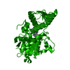
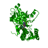
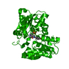
| Entry | Database: PDB / ID: 4ho1 | ||||||
|---|---|---|---|---|---|---|---|
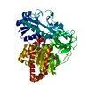
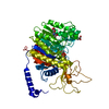
| Title | The Crystal structure of Haemophilus influenzae O-acetylserine sulfhydrylase at 1.85A resolution | ||||||
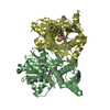
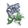
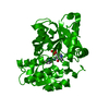
 Components Components | Cysteine synthase | ||||||
 Keywords Keywords | TRANSFERASE / alpha/beta fold / Rossmann fold CysK / Enzyme / Rossmann fold / catalytic activity / cysteine synthase activity / transferase activity / serine acetyl transferase | ||||||
| Function / homology |  Function and homology informationcysteine synthase / cystathionine beta-synthase activity / cysteine synthase activity / L-cysteine desulfhydrase activity / cysteine biosynthetic process from serine / cytoplasm Function and homology informationcysteine synthase / cystathionine beta-synthase activity / cysteine synthase activity / L-cysteine desulfhydrase activity / cysteine biosynthetic process from serine / cytoplasmSimilarity search - Function | ||||||
| Biological species |  Haemophilus influenzae (bacteria) Haemophilus influenzae (bacteria) | ||||||
| Method | X-RAY DIFFRACTION / MOLECULAR REPLACEMENT / Resolution: 1.856 Å | ||||||
 Authors Authors | Banerjee, S. / Singh, A.K. / Kumaran, S. | ||||||
 Citation Citation | Journal: TO BE PUBLISHED Title: The Crystal structure of Haemophilus influenzae O-acetylserine sulfhydrylase at 1.85A resolution Authors: Banerjee, S. / Singh, A.K. / Kumaran, S. | ||||||
| History |
|
- Structure visualization
| Structure viewer | Molecule: MolmilJmol/JSmol |
|---|
- Downloads & links
-Download
| PDBx/mmCIF format | 4ho1.cif.gz | 76.1 KB | Display | PDBx/mmCIF format |
|---|---|---|---|---|
| PDB format | pdb4ho1.ent.gz | 54.9 KB | Display | PDB format |
| PDBx/mmJSON format | 4ho1.json.gz | Tree view | PDBx/mmJSON format | |
| Others |  Other downloads Other downloads |
-Validation report
| Arichive directory | https://data.pdbj.org/pub/pdb/validation_reports/ho/4ho1ftp://data.pdbj.org/pub/pdb/validation_reports/ho/4ho1 | HTTPS FTP |
|---|
-Related structure data
| Related structure data | 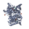 1y7lS S: Starting model for refinement |
|---|---|
| Similar structure data |

-Links
 PDBj
PDBj
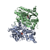
- Assembly
| Deposited unit | 
| ||||||||
|---|---|---|---|---|---|---|---|---|---|
| 1 | 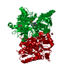
| ||||||||
| Unit cell |
| ||||||||
| Components on special symmetry positions |
|
-Components
| #1: Protein | / CSase / O-acetylserine (thiol)-lyase / OAS-TL / O-acetylserine sulfhydrylase Mass: 33651.465 Da / Num. of mol.: 1 / Fragment: O-acetylserine sulfhydrylase Source method: isolated from a genetically manipulated source Source: (gene. exp.) Haemophilus influenzae (bacteria) / Strain: ATCC 51907 / Gene: cysK, HI_1103 / Plasmid: pET28A / Production host: Escherichia coli (E. coli) / Strain (production host): K-12 / References: UniProt: P45040, cysteine synthase | ||
|---|---|---|---|
| #2: Chemical | ChemComp-GOL / Glycerol  Mass: 92.094 Da / Num. of mol.: 1 / Source method: obtained synthetically / Formula: C3H8O3 Mass: 92.094 Da / Num. of mol.: 1 / Source method: obtained synthetically / Formula: C3H8O3 | ||
| #3: Chemical | Ethylene glycol  Mass: 62.068 Da / Num. of mol.: 2 / Source method: obtained synthetically / Formula: C2H6O2 Mass: 62.068 Da / Num. of mol.: 2 / Source method: obtained synthetically / Formula: C2H6O2#4: Water | ChemComp-HOH / | Water Mass: 18.015 Da / Num. of mol.: 135 / Source method: isolated from a natural source / Formula: H2O Mass: 18.015 Da / Num. of mol.: 135 / Source method: isolated from a natural source / Formula: H2O |
-Experimental details
-Experiment
| Experiment | Method: X-RAY DIFFRACTION / Number of used crystals: 1 |
|---|
- Sample preparation
| Crystal | Density Matthews: 2.19 Å3/Da / Density % sol: 43.77 % |
|---|---|
| Crystal grow | Temperature: 292 K / Method: vapor diffusion, sitting drop / pH: 8.5 Details: 0.2M Magnesium chloride,0.1M tris HCL pH 8.5, 30% w/v PEG 4000 , VAPOR DIFFUSION, SITTING DROP, temperature 292K |
-Data collection
| Diffraction | Mean temperature: 100 K | |||||||||||||||
|---|---|---|---|---|---|---|---|---|---|---|---|---|---|---|---|---|
| Diffraction source | Source: ROTATING ANODE / Type: RIGAKU MICROMAX-007 HF / Wavelength: 1.5418 Å | |||||||||||||||
| Detector | Type: MAR scanner 345 mm plate / Detector: IMAGE PLATE / Date: Apr 6, 2010 | |||||||||||||||
| Radiation | Monochromator: Graphite polar / Protocol: SINGLE WAVELENGTH / Monochromatic (M) / Laue (L): M / Scattering type: x-ray | |||||||||||||||
| Radiation wavelength | Wavelength: 1.5418 Å / Relative weight: 1 | |||||||||||||||
| Reflection twin |
| |||||||||||||||
| Reflection | Resolution: 1.856→79.75 Å / Num. all: 24841 / Num. obs: 24451 / % possible obs: 97.7 % / Observed criterion σ(F): 2 / Observed criterion σ(I): 2 / Redundancy: 7.4 % / Rmerge(I) obs: 0.044 / Net I/σ(I): 34.81 |
- Processing
| Software |
| ||||||||||||||||||||||||||||||||||||||||||||||||||||||||||||||||||||||||||||||||||||||||||||||||||||||||||||||||||||||||||||||||||||||||||||||||||||||||||||||||||||||||||||||||||||||
|---|---|---|---|---|---|---|---|---|---|---|---|---|---|---|---|---|---|---|---|---|---|---|---|---|---|---|---|---|---|---|---|---|---|---|---|---|---|---|---|---|---|---|---|---|---|---|---|---|---|---|---|---|---|---|---|---|---|---|---|---|---|---|---|---|---|---|---|---|---|---|---|---|---|---|---|---|---|---|---|---|---|---|---|---|---|---|---|---|---|---|---|---|---|---|---|---|---|---|---|---|---|---|---|---|---|---|---|---|---|---|---|---|---|---|---|---|---|---|---|---|---|---|---|---|---|---|---|---|---|---|---|---|---|---|---|---|---|---|---|---|---|---|---|---|---|---|---|---|---|---|---|---|---|---|---|---|---|---|---|---|---|---|---|---|---|---|---|---|---|---|---|---|---|---|---|---|---|---|---|---|---|---|---|
| Refinement | Method to determine structure: MOLECULAR REPLACEMENT Starting model: PDB ENTRY 1Y7L Resolution: 1.856→35.67 Å / Cor.coef. Fo:Fc: 0.969 / Cor.coef. Fo:Fc free: 0.961 / SU B: 2.851 / SU ML: 0.087 / Isotropic thermal model: Isotropic / Cross valid method: THROUGHOUT / ESU R: 0.024 / ESU R Free: 0.021 / Stereochemistry target values: MAXIMUM LIKELIHOOD / Details: HYDROGENS HAVE BEEN ADDED IN THE RIDING POSITIONS
| ||||||||||||||||||||||||||||||||||||||||||||||||||||||||||||||||||||||||||||||||||||||||||||||||||||||||||||||||||||||||||||||||||||||||||||||||||||||||||||||||||||||||||||||||||||||
| Solvent computation | Ion probe radii: 0.8 Å / Shrinkage radii: 0.8 Å / VDW probe radii: 1.2 Å / Solvent model: MASK | ||||||||||||||||||||||||||||||||||||||||||||||||||||||||||||||||||||||||||||||||||||||||||||||||||||||||||||||||||||||||||||||||||||||||||||||||||||||||||||||||||||||||||||||||||||||
| Displacement parameters | Biso mean: 32.322 Å2
| ||||||||||||||||||||||||||||||||||||||||||||||||||||||||||||||||||||||||||||||||||||||||||||||||||||||||||||||||||||||||||||||||||||||||||||||||||||||||||||||||||||||||||||||||||||||
| Refinement step | Cycle: LAST / Resolution: 1.856→35.67 Å
| ||||||||||||||||||||||||||||||||||||||||||||||||||||||||||||||||||||||||||||||||||||||||||||||||||||||||||||||||||||||||||||||||||||||||||||||||||||||||||||||||||||||||||||||||||||||
| Refine LS restraints |
|