 Movie
Movie Controller

[English] 日本語
 Yorodumi
- PDB-7efp: Structure of SARS-CoV-2 spike receptor-binding domain in complex ... -
+ Open data

- Basic information
| Entry | Database: PDB / ID: 7efp | ||||||
|---|---|---|---|---|---|---|---|
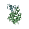
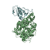
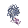
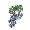
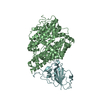
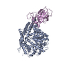
| Title | Structure of SARS-CoV-2 spike receptor-binding domain in complex with high affinity ACE2 mutant (S19W,N330Y) | ||||||
 Components Components |
| ||||||
 Keywords Keywords |  VIRAL PROTEIN / receptor-binding domain / angiotensin converting enzyme 2 VIRAL PROTEIN / receptor-binding domain / angiotensin converting enzyme 2 | ||||||
| Function / homology |  Function and homology information Function and homology informationpositive regulation of amino acid transport / angiotensin-converting enzyme 2 / positive regulation of L-proline import across plasma membrane / Hydrolases; Acting on peptide bonds (peptidases); Metallocarboxypeptidases / angiotensin-mediated drinking behavior / tryptophan transport / positive regulation of gap junction assembly / regulation of systemic arterial blood pressure by renin-angiotensin / regulation of vasoconstriction / regulation of cardiac conduction ...positive regulation of amino acid transport / angiotensin-converting enzyme 2 / positive regulation of L-proline import across plasma membrane / Hydrolases; Acting on peptide bonds (peptidases); Metallocarboxypeptidases / angiotensin-mediated drinking behavior / tryptophan transport / positive regulation of gap junction assembly / regulation of systemic arterial blood pressure by renin-angiotensin / regulation of vasoconstriction / regulation of cardiac conduction / peptidyl-dipeptidase activity / angiotensin maturation / maternal process involved in female pregnancy / Metabolism of Angiotensinogen to Angiotensins / metallocarboxypeptidase activity / Attachment and Entry / carboxypeptidase activity / negative regulation of signaling receptor activity / positive regulation of cardiac muscle contraction / regulation of cytokine production / viral life cycle / blood vessel diameter maintenance / regulation of transmembrane transporter activity / brush border membrane / negative regulation of smooth muscle cell proliferation / cilium / negative regulation of ERK1 and ERK2 cascade / endocytic vesicle membrane / metallopeptidase activity / positive regulation of reactive oxygen species metabolic process / virus receptor activity / regulation of cell population proliferation / regulation of inflammatory response / Maturation of spike protein / viral translation / Translation of Structural Proteins / Virion Assembly and Release / host cell surface / host extracellular space / endopeptidase activity / suppression by virus of host tetherin activity / Induction of Cell-Cell Fusion / structural constituent of virion / Potential therapeutics for SARS / entry receptor-mediated virion attachment to host cell / host cell endoplasmic reticulum-Golgi intermediate compartment membrane / receptor-mediated endocytosis of virus by host cell / Attachment and Entry / membrane fusion / positive regulation of viral entry into host cell / receptor-mediated virion attachment to host cell / receptor ligand activity / host cell surface receptor binding / symbiont entry into host cell / membrane raft / apical plasma membrane / fusion of virus membrane with host plasma membrane / endoplasmic reticulum lumen / fusion of virus membrane with host endosome membrane / viral envelope / symbiont-mediated suppression of host type I interferon-mediated signaling pathway / virion attachment to host cell / SARS-CoV-2 activates/modulates innate and adaptive immune responses / host cell plasma membrane / virion membrane / cell surface / extracellular space / extracellular exosome / zinc ion binding / extracellular region / membrane / identical protein binding / plasma membraneSimilarity search - Function | ||||||
| Biological species |  Homo sapiens (human) Homo sapiens (human)  Severe acute respiratory syndrome coronavirus 2 Severe acute respiratory syndrome coronavirus 2 | ||||||
| Method | X-RAY DIFFRACTION / SYNCHROTRON / MOLECULAR REPLACEMENT / Resolution: 2.698 Å | ||||||
 Authors Authors | Lu, G.W. / Ye, F. / Lin, X. | ||||||
 Citation Citation | Journal: Signal Transduct Target Ther / Year: 2021 Title: S19W, T27W, and N330Y mutations in ACE2 enhance SARS-CoV-2 S-RBD binding toward both wild-type and antibody-resistant viruses and its molecular basis. Authors: Ye, F. / Lin, X. / Chen, Z. / Yang, F. / Lin, S. / Yang, J. / Chen, H. / Sun, H. / Wang, L. / Wen, A. / Zhang, X. / Dai, Y. / Cao, Y. / Yang, J. / Shen, G. / Yang, L. / Li, J. / Wang, Z. / ...Authors: Ye, F. / Lin, X. / Chen, Z. / Yang, F. / Lin, S. / Yang, J. / Chen, H. / Sun, H. / Wang, L. / Wen, A. / Zhang, X. / Dai, Y. / Cao, Y. / Yang, J. / Shen, G. / Yang, L. / Li, J. / Wang, Z. / Wang, W. / Wei, X. / Lu, G. | ||||||
| History |
|
- Structure visualization
| Structure viewer | Molecule: MolmilJmol/JSmol |
|---|
- Downloads & links
-Download
| PDBx/mmCIF format | 7efp.cif.gz | 338 KB | Display | PDBx/mmCIF format |
|---|---|---|---|---|
| PDB format | pdb7efp.ent.gz | 273.4 KB | Display | PDB format |
| PDBx/mmJSON format | 7efp.json.gz | Tree view | PDBx/mmJSON format | |
| Others |  Other downloads Other downloads |
-Validation report
| Arichive directory | https://data.pdbj.org/pub/pdb/validation_reports/ef/7efpftp://data.pdbj.org/pub/pdb/validation_reports/ef/7efp | HTTPS FTP |
|---|
-Related structure data
| Related structure data | 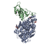 7efrC 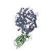 6lzgS S: Starting model for refinement C: citing same article ( |
|---|---|
| Similar structure data |


-Links
 PDBj
PDBj


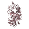
- Assembly
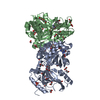
| Deposited unit | 
| ||||||||
|---|---|---|---|---|---|---|---|---|---|
| 1 |
| ||||||||
| Unit cell |
|
-Components
| #1: Protein | Mass: 69488.039 Da / Num. of mol.: 1 / Mutation: S19W, N330Y Source method: isolated from a genetically manipulated source Source: (gene. exp.) Homo sapiens (human) / Gene: ACE2, UNQ868/PRO1885 / Production host:  Trichoplusia ni (cabbage looper) / References: UniProt: Q9BYF1 Trichoplusia ni (cabbage looper) / References: UniProt: Q9BYF1 | ||||
|---|---|---|---|---|---|
| #2: Protein | Spike protein / S glycoprotein / E2 / Peplomer protein Mass: 24501.592 Da / Num. of mol.: 1 / Fragment: UNP 320-537 Source method: isolated from a genetically manipulated source Source: (gene. exp.) Severe acute respiratory syndrome coronavirus 2Gene: S, 2 / Production host: Trichoplusia ni (cabbage looper) / References: UniProt: P0DTC2 | ||||
| #3: Sugar | ChemComp-NAG / N-Acetylglucosamine  Type: D-saccharide, beta linking / Mass: 221.208 Da / Num. of mol.: 5 / Source method: obtained synthetically / Formula: C8H15NO6 Type: D-saccharide, beta linking / Mass: 221.208 Da / Num. of mol.: 5 / Source method: obtained synthetically / Formula: C8H15NO6#4: Water | ChemComp-HOH / | Water Mass: 18.015 Da / Num. of mol.: 58 / Source method: isolated from a natural source / Formula: H2O Mass: 18.015 Da / Num. of mol.: 58 / Source method: isolated from a natural source / Formula: H2OHas ligand of interest | N | |
-Experimental details
-Experiment
| Experiment | Method: X-RAY DIFFRACTION / Number of used crystals: 1 |
|---|
- Sample preparation
| Crystal | Density Matthews: 4.04 Å3/Da / Density % sol: 69.57 % |
|---|---|
| Crystal grow | Temperature: 291 K / Method: vapor diffusion, sitting drop / pH: 6.5 Details: 1.6 M Ammonium sulfate, 0.1 M MES pH6.5, 10 % v/v 1,4-Dioxane |
-Data collection
| Diffraction | Mean temperature: 100 K / Serial crystal experiment: N |
|---|---|
| Diffraction source | Source: SYNCHROTRON / Site: SSRF  / Beamline: BL18U1 / Wavelength: 0.97915 Å / Beamline: BL18U1 / Wavelength: 0.97915 Å |
| Detector | Type: DECTRIS PILATUS3 S 6M / Detector: PIXEL / Date: Jul 23, 2020 |
| Radiation | Protocol: SINGLE WAVELENGTH / Monochromatic (M) / Laue (L): M / Scattering type: x-ray |
| Radiation wavelength | Wavelength: 0.97915 Å / Relative weight: 1 |
| Reflection | Resolution: 2.69→50 Å / Num. obs: 42978 / % possible obs: 100 % / Redundancy: 12.3 % / Rmerge(I) obs: 0.155 / Net I/σ(I): 19.6 |
| Reflection shell | Resolution: 2.69→2.8 Å / Rmerge(I) obs: 1.297 / Num. unique obs: 4234 |
- Processing
| Software |
| ||||||||||||||||||||||||||||||||||||||||||||||||||||||||||||||||||||||||||||||||||||||||||||||||
|---|---|---|---|---|---|---|---|---|---|---|---|---|---|---|---|---|---|---|---|---|---|---|---|---|---|---|---|---|---|---|---|---|---|---|---|---|---|---|---|---|---|---|---|---|---|---|---|---|---|---|---|---|---|---|---|---|---|---|---|---|---|---|---|---|---|---|---|---|---|---|---|---|---|---|---|---|---|---|---|---|---|---|---|---|---|---|---|---|---|---|---|---|---|---|---|---|---|
| Refinement | Method to determine structure: MOLECULAR REPLACEMENT Starting model: 6LZG Resolution: 2.698→25.922 Å / SU ML: 0.35 / Cross valid method: THROUGHOUT / σ(F): 1.35 / Phase error: 24.59 / Stereochemistry target values: ML
| ||||||||||||||||||||||||||||||||||||||||||||||||||||||||||||||||||||||||||||||||||||||||||||||||
| Solvent computation | Shrinkage radii: 0.9 Å / VDW probe radii: 1.11 Å / Solvent model: FLAT BULK SOLVENT MODEL | ||||||||||||||||||||||||||||||||||||||||||||||||||||||||||||||||||||||||||||||||||||||||||||||||
| Displacement parameters | Biso max: 168.03 Å2 / Biso mean: 58.4597 Å2 / Biso min: 34.23 Å2 | ||||||||||||||||||||||||||||||||||||||||||||||||||||||||||||||||||||||||||||||||||||||||||||||||
| Refinement step | Cycle: final / Resolution: 2.698→25.922 Å
| ||||||||||||||||||||||||||||||||||||||||||||||||||||||||||||||||||||||||||||||||||||||||||||||||
| LS refinement shell | Refine-ID: X-RAY DIFFRACTION / Rfactor Rfree error: 0
| ||||||||||||||||||||||||||||||||||||||||||||||||||||||||||||||||||||||||||||||||||||||||||||||||
| Refinement TLS params. | Method: refined / Origin x: -34.2562 Å / Origin y: -16.3506 Å / Origin z: -38.7675 Å
| ||||||||||||||||||||||||||||||||||||||||||||||||||||||||||||||||||||||||||||||||||||||||||||||||
| Refinement TLS group |
|
