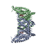
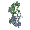
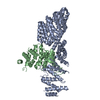
 Movie
Movie Controller

[English] 日本語
 Yorodumi
- PDB-7e9s: Archaeal oligosaccharyltransferase AglB from Archaeoglobus fulgid... -
+ Open data

- Basic information
| Entry | Database: PDB / ID: 7e9s | |||||||||
|---|---|---|---|---|---|---|---|---|---|---|
| Title | Archaeal oligosaccharyltransferase AglB from Archaeoglobus fulgidus in complex with an inhibitory peptide and a dolichol-phosphate | |||||||||
 Components Components |
| |||||||||
 Keywords Keywords |  TRANSFERASE / protein n-glycosylation / ternary complex / sequon-containing peptide / dolichol-phosphate TRANSFERASE / protein n-glycosylation / ternary complex / sequon-containing peptide / dolichol-phosphate | |||||||||
| Function / homology |  Function and homology information Function and homology informationdolichyl-phosphooligosaccharide-protein glycotransferase / oligosaccharyl transferase activity / protein glycosylation / metal ion binding / plasma membraneSimilarity search - Function | |||||||||
| Biological species |   Archaeoglobus fulgidus DSM 4304 (archaea) Archaeoglobus fulgidus DSM 4304 (archaea)synthetic construct (others) | |||||||||
| Method | X-RAY DIFFRACTION / SYNCHROTRON / MOLECULAR REPLACEMENT / Resolution: 2.7 Å | |||||||||
 Authors Authors | Taguchi, Y. / Hirata, K. / Kohda, D. | |||||||||
| Funding support |  Japan, 2items Japan, 2items
| |||||||||
 Citation Citation | Journal: Commun Biol / Year: 2021 Title: The structure of an archaeal oligosaccharyltransferase provides insight into the strict exclusion of proline from the N-glycosylation sequon. Authors: Taguchi, Y. / Yamasaki, T. / Ishikawa, M. / Kawasaki, Y. / Yukimura, R. / Mitani, M. / Hirata, K. / Kohda, D. | |||||||||
| History |
|
- Structure visualization
| Structure viewer | Molecule: MolmilJmol/JSmol |
|---|
- Downloads & links
-Download
| PDBx/mmCIF format | 7e9s.cif.gz | 439 KB | Display | PDBx/mmCIF format |
|---|---|---|---|---|
| PDB format | pdb7e9s.ent.gz | 298.3 KB | Display | PDB format |
| PDBx/mmJSON format | 7e9s.json.gz | Tree view | PDBx/mmJSON format | |
| Others |  Other downloads Other downloads |
-Validation report
| Arichive directory | https://data.pdbj.org/pub/pdb/validation_reports/e9/7e9sftp://data.pdbj.org/pub/pdb/validation_reports/e9/7e9s | HTTPS FTP |
|---|
-Related structure data
| Related structure data |  5gmyS S: Starting model for refinement |
|---|---|
| Similar structure data |


-Links
 PDBj
PDBj
- Assembly
| Deposited unit | 
| ||||||||||||
|---|---|---|---|---|---|---|---|---|---|---|---|---|---|
| 1 |
| ||||||||||||
| Unit cell |
|
-Components
-Protein / Protein/peptide , 2 types, 2 molecules AB
| #1: Protein | Mass: 99159.875 Da / Num. of mol.: 1 / Mutation: G617C Source method: isolated from a genetically manipulated source Source: (gene. exp.) Archaeoglobus fulgidus DSM 4304 (archaea)Strain: DSM 4304 / Gene: aglB3, AF_0380 / Plasmid: pET-52b(+) / Production host:  Escherichia coli BL21(DE3) (bacteria) / Strain (production host): BL21(DE3) / Variant (production host): C43 Escherichia coli BL21(DE3) (bacteria) / Strain (production host): BL21(DE3) / Variant (production host): C43References: UniProt: O29867, dolichyl-phosphooligosaccharide-protein glycotransferase |
|---|---|
| #2: Protein/peptide | Mass: 1068.227 Da / Num. of mol.: 1 / Source method: obtained synthetically / Source: (synth.) synthetic construct (others) |
-Non-polymers , 5 types, 27 molecules 


| #3: Chemical | ChemComp-MN /  Mass: 54.938 Da / Num. of mol.: 1 / Source method: obtained synthetically / Formula: Mn Mass: 54.938 Da / Num. of mol.: 1 / Source method: obtained synthetically / Formula: Mn | ||||
|---|---|---|---|---|---|
| #4: Chemical | ChemComp-J06 / [( Mass: 923.463 Da / Num. of mol.: 1 / Source method: obtained synthetically / Formula: C60H107O4P / Feature type: SUBJECT OF INVESTIGATION Mass: 923.463 Da / Num. of mol.: 1 / Source method: obtained synthetically / Formula: C60H107O4P / Feature type: SUBJECT OF INVESTIGATION | ||||
| #5: Chemical | ChemComp-PEG / Diethylene glycol Mass: 106.120 Da / Num. of mol.: 5 / Source method: obtained synthetically / Formula: C4H10O3 Mass: 106.120 Da / Num. of mol.: 5 / Source method: obtained synthetically / Formula: C4H10O3#6: Chemical | ChemComp-7E8 / (  Mass: 300.434 Da / Num. of mol.: 4 / Source method: obtained synthetically / Formula: C17H32O4 Mass: 300.434 Da / Num. of mol.: 4 / Source method: obtained synthetically / Formula: C17H32O4#7: Water | ChemComp-HOH / | WaterMass: 18.015 Da / Num. of mol.: 16 / Source method: isolated from a natural source / Formula: H2O |
-Details
| Has ligand of interest | Y |
|---|---|
| Nonpolymer details | We used TMR(CARBOXYTETRAMETHYLRHODAMINE) to stand for tetramethylrhodamine (RHO), which is a ...We used TMR(CARBOXYTET |
-Experimental details
-Experiment
| Experiment | Method: X-RAY DIFFRACTION / Number of used crystals: 1 |
|---|
- Sample preparation
| Crystal | Density Matthews: 2.66 Å3/Da / Density % sol: 53.84 % Description: The microcrystals were needle-shaped with a length greater than 100 micro m and a width/thickness less than 5 micro m. |
|---|---|
| Crystal grow | Temperature: 293 K / Method: lipidic cubic phase / pH: 6 Details: 50 mM NaCl, 0.1 M Na-citrate, pH 6.0, 18-22 % PEG400 |
-Data collection
| Diffraction | Mean temperature: 100 K / Ambient temp details: cryo N2 stream / Serial crystal experiment: N |
|---|---|
| Diffraction source | Source: SYNCHROTRON / Site: SPring-8 / Beamline: BL32XU / Wavelength: 1 Å |
| Detector | Type: DECTRIS EIGER X 9M / Detector: PIXEL / Date: Feb 20, 2019 Details: micro-focused beam of 10 micro m x 15 micro m (horizontal x vertical) |
| Radiation | Protocol: SINGLE WAVELENGTH / Monochromatic (M) / Laue (L): M / Scattering type: x-ray |
| Radiation wavelength | Wavelength: 1 Å / Relative weight: 1 |
| Reflection | Resolution: 2.7→49.4 Å / Num. obs: 30677 / % possible obs: 100 % / Redundancy: 79.5 % / Biso Wilson estimate: 70.16 Å2 / CC1/2: 0.998 / Rmerge(I) obs: 0.432 / Rrim(I) all: 0.435 / Net I/σ(I): 13.6 |
| Reflection shell | Resolution: 2.7→2.8 Å / Redundancy: 76.9 % / Rmerge(I) obs: 8.718 / Mean I/σ(I) obs: 1.3 / Num. unique obs: 3054 / CC1/2: 0.756 / Rrim(I) all: 8.776 / % possible all: 99.9 |
- Processing
| Software |
| ||||||||||||||||||||||||||||||||||||||||||||||||||||||||||||||||||||||||||||||||||||||||||||||||||||
|---|---|---|---|---|---|---|---|---|---|---|---|---|---|---|---|---|---|---|---|---|---|---|---|---|---|---|---|---|---|---|---|---|---|---|---|---|---|---|---|---|---|---|---|---|---|---|---|---|---|---|---|---|---|---|---|---|---|---|---|---|---|---|---|---|---|---|---|---|---|---|---|---|---|---|---|---|---|---|---|---|---|---|---|---|---|---|---|---|---|---|---|---|---|---|---|---|---|---|---|---|---|
| Refinement | Method to determine structure: MOLECULAR REPLACEMENT Starting model: 5GMY Resolution: 2.7→24.84 Å / SU ML: 0.3262 / Cross valid method: FREE R-VALUE / σ(F): 1.35 / Phase error: 27.7132 Stereochemistry target values: GeoStd + Monomer Library + CDL v1.2
| ||||||||||||||||||||||||||||||||||||||||||||||||||||||||||||||||||||||||||||||||||||||||||||||||||||
| Solvent computation | Shrinkage radii: 0.9 Å / VDW probe radii: 1.11 Å / Solvent model: FLAT BULK SOLVENT MODEL | ||||||||||||||||||||||||||||||||||||||||||||||||||||||||||||||||||||||||||||||||||||||||||||||||||||
| Displacement parameters | Biso mean: 79 Å2 | ||||||||||||||||||||||||||||||||||||||||||||||||||||||||||||||||||||||||||||||||||||||||||||||||||||
| Refinement step | Cycle: LAST / Resolution: 2.7→24.84 Å
| ||||||||||||||||||||||||||||||||||||||||||||||||||||||||||||||||||||||||||||||||||||||||||||||||||||
| Refine LS restraints |
| ||||||||||||||||||||||||||||||||||||||||||||||||||||||||||||||||||||||||||||||||||||||||||||||||||||
| LS refinement shell |
| ||||||||||||||||||||||||||||||||||||||||||||||||||||||||||||||||||||||||||||||||||||||||||||||||||||
| Refinement TLS params. | Method: refined / Refine-ID: X-RAY DIFFRACTION
| ||||||||||||||||||||||||||||||||||||||||||||||||||||||||||||||||||||||||||||||||||||||||||||||||||||
| Refinement TLS group | Refine-ID: X-RAY DIFFRACTION
|
