 Movie
Movie Controller


+ Open data

- Basic information
| Entry | Database: PDB / ID: 7e7f | |||||||||
|---|---|---|---|---|---|---|---|---|---|---|
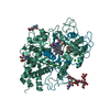
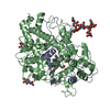
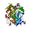
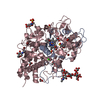
| Title | Human CYP11B1 mutant in complex with metyrapone | |||||||||
 Components Components | Cytochrome P450 11B1, mitochondrial | |||||||||
 Keywords Keywords |  OXIDOREDUCTASE / P450 OXIDOREDUCTASE / P450 | |||||||||
| Function / homology |  Function and homology information Function and homology informationDefective CYP11B1 causes AH4 / steroid 11beta-monooxygenase / steroid 11-beta-monooxygenase activity / corticosterone 18-monooxygenase activity / cortisol metabolic process / aldosterone biosynthetic process / cortisol biosynthetic process / glucocorticoid biosynthetic process / Glucocorticoid biosynthesis / sterol metabolic process ...Defective CYP11B1 causes AH4 / steroid 11beta-monooxygenase / steroid 11-beta-monooxygenase activity / corticosterone 18-monooxygenase activity / cortisol metabolic process / aldosterone biosynthetic process / cortisol biosynthetic process / glucocorticoid biosynthetic process / Glucocorticoid biosynthesis / sterol metabolic process / cellular response to potassium ion / C21-steroid hormone biosynthetic process / cellular response to peptide hormone stimulus / Endogenous sterols / cellular response to hormone stimulus / cholesterol metabolic process / regulation of blood pressure / glucose homeostasis / mitochondrial inner membrane / immune response / iron ion binding / heme binding / mitochondrionSimilarity search - Function | |||||||||
| Biological species |  Homo sapiens (human) Homo sapiens (human) | |||||||||
| Method | X-RAY DIFFRACTION / SYNCHROTRON / MOLECULAR REPLACEMENT / Resolution: 1.4 Å | |||||||||
| Model details | Mitocondrial cytochrome P450 11B1 | |||||||||
 Authors Authors | Mukai, K. / Sugimoto, H. / Reiko, S. / Matsuura, T. / Hishiki, T. / Kagawa, N. | |||||||||
| Funding support |  Japan, 2items Japan, 2items
| |||||||||
 Citation Citation | Journal: Curr Res Struct Biol / Year: 2021 Title: Spatially restricted substrate-binding site of cortisol-synthesizing CYP11B1 limits multiple hydroxylations and hinders aldosterone synthesis. Authors: Mukai, K. / Sugimoto, H. / Kamiya, K. / Suzuki, R. / Matsuura, T. / Hishiki, T. / Shimada, H. / Shiro, Y. / Suematsu, M. / Kagawa, N. | |||||||||
| History |
|
- Structure visualization
| Structure viewer | Molecule: MolmilJmol/JSmol |
|---|
- Downloads & links
-Download
| PDBx/mmCIF format | 7e7f.cif.gz | 236.6 KB | Display | PDBx/mmCIF format |
|---|---|---|---|---|
| PDB format | pdb7e7f.ent.gz | 185.1 KB | Display | PDB format |
| PDBx/mmJSON format | 7e7f.json.gz | Tree view | PDBx/mmJSON format | |
| Others |  Other downloads Other downloads |
-Validation report
| Arichive directory | https://data.pdbj.org/pub/pdb/validation_reports/e7/7e7fftp://data.pdbj.org/pub/pdb/validation_reports/e7/7e7f | HTTPS FTP |
|---|
-Related structure data
| Related structure data |  4dvqS S: Starting model for refinement |
|---|---|
| Similar structure data |


-Links
 PDBj
PDBj

- Assembly
| Deposited unit | 
| ||||||||
|---|---|---|---|---|---|---|---|---|---|
| 1 |
| ||||||||
| Unit cell |
|
-Components
-Protein , 1 types, 1 molecules A
| #1: Protein | Mass: 55821.336 Da / Num. of mol.: 1 / Fragment: heme binding protein / Mutation: W49R,L50N,W53R,W56R,L244N,W247N Source method: isolated from a genetically manipulated source Source: (gene. exp.) Homo sapiens (human) / Gene: CYP11B1, S11BH / Plasmid: pET-17b / Production host:  Escherichia coli BL21(DE3) (bacteria) / References: UniProt: P15538, steroid 11beta-monooxygenase Escherichia coli BL21(DE3) (bacteria) / References: UniProt: P15538, steroid 11beta-monooxygenase |
|---|
-Non-polymers , 5 types, 424 molecules 


| #2: Chemical | ChemComp-HEM / Heme B Mass: 616.487 Da / Num. of mol.: 1 / Source method: obtained synthetically / Formula: C34H32FeN4O4 Mass: 616.487 Da / Num. of mol.: 1 / Source method: obtained synthetically / Formula: C34H32FeN4O4 | ||||
|---|---|---|---|---|---|
| #3: Chemical | ChemComp-MYT / Metyrapone Mass: 226.274 Da / Num. of mol.: 1 / Source method: obtained synthetically / Formula: C14H14N2O / Feature type: SUBJECT OF INVESTIGATION / Comment: medication, inhibitor*YM Mass: 226.274 Da / Num. of mol.: 1 / Source method: obtained synthetically / Formula: C14H14N2O / Feature type: SUBJECT OF INVESTIGATION / Comment: medication, inhibitor*YM | ||||
| #4: Chemical | Cholic acid Mass: 408.571 Da / Num. of mol.: 2 / Source method: obtained synthetically / Formula: C24H40O5 Mass: 408.571 Da / Num. of mol.: 2 / Source method: obtained synthetically / Formula: C24H40O5#5: Chemical | Glycerol Mass: 92.094 Da / Num. of mol.: 2 / Source method: obtained synthetically / Formula: C3H8O3 Mass: 92.094 Da / Num. of mol.: 2 / Source method: obtained synthetically / Formula: C3H8O3#6: Water | ChemComp-HOH / | WaterMass: 18.015 Da / Num. of mol.: 418 / Source method: isolated from a natural source / Formula: H2O |
-Details
| Has ligand of interest | Y |
|---|
-Experimental details
-Experiment
| Experiment | Method: X-RAY DIFFRACTION / Number of used crystals: 1 |
|---|
- Sample preparation
| Crystal | Density Matthews: 2.44 Å3/Da / Density % sol: 49.56 % |
|---|---|
| Crystal grow | Temperature: 277 K / Method: vapor diffusion, sitting drop / pH: 7.4 Details: 100mM potassium phosphate, 150mM NaCl, 4% w/v PEG 3350 |
-Data collection
| Diffraction | Mean temperature: 100 K / Serial crystal experiment: N | ||||||||||||||||||||||||||||||||||||||||||||||||||||||||||||||||||||||||||||||||||||||||||||||||||||
|---|---|---|---|---|---|---|---|---|---|---|---|---|---|---|---|---|---|---|---|---|---|---|---|---|---|---|---|---|---|---|---|---|---|---|---|---|---|---|---|---|---|---|---|---|---|---|---|---|---|---|---|---|---|---|---|---|---|---|---|---|---|---|---|---|---|---|---|---|---|---|---|---|---|---|---|---|---|---|---|---|---|---|---|---|---|---|---|---|---|---|---|---|---|---|---|---|---|---|---|---|---|
| Diffraction source | Source: SYNCHROTRON / Site: SPring-8 / Beamline: BL41XU / Wavelength: 1 Å | ||||||||||||||||||||||||||||||||||||||||||||||||||||||||||||||||||||||||||||||||||||||||||||||||||||
| Detector | Type: DECTRIS EIGER X 16M / Detector: PIXEL / Date: Feb 14, 2018 / Details: mirrors | ||||||||||||||||||||||||||||||||||||||||||||||||||||||||||||||||||||||||||||||||||||||||||||||||||||
| Radiation | Monochromator: Si(111) / Protocol: SINGLE WAVELENGTH / Monochromatic (M) / Laue (L): M / Scattering type: x-ray | ||||||||||||||||||||||||||||||||||||||||||||||||||||||||||||||||||||||||||||||||||||||||||||||||||||
| Radiation wavelength | Wavelength: 1 Å / Relative weight: 1 | ||||||||||||||||||||||||||||||||||||||||||||||||||||||||||||||||||||||||||||||||||||||||||||||||||||
| Reflection | Resolution: 1.4→47.02 Å / Num. obs: 105301 / % possible obs: 97.6 % / Redundancy: 17.249 % / Biso Wilson estimate: 30.218 Å2 / CC1/2: 1 / Rmerge(I) obs: 0.045 / Rrim(I) all: 0.046 / Χ2: 1.036 / Net I/σ(I): 29.34 / Num. measured all: 1816384 / Scaling rejects: 2235 | ||||||||||||||||||||||||||||||||||||||||||||||||||||||||||||||||||||||||||||||||||||||||||||||||||||
| Reflection shell | Diffraction-ID: 1
|
- Processing
| Software |
| |||||||||||||||||||||||||||||||||||||||||||||||||||||||||||||||||
|---|---|---|---|---|---|---|---|---|---|---|---|---|---|---|---|---|---|---|---|---|---|---|---|---|---|---|---|---|---|---|---|---|---|---|---|---|---|---|---|---|---|---|---|---|---|---|---|---|---|---|---|---|---|---|---|---|---|---|---|---|---|---|---|---|---|---|
| Refinement | Method to determine structure: MOLECULAR REPLACEMENT Starting model: 4DVQ Resolution: 1.4→47.02 Å / Cor.coef. Fo:Fc: 0.978 / Cor.coef. Fo:Fc free: 0.975 / SU B: 1.969 / SU ML: 0.034 / SU R Cruickshank DPI: 0.0544 / Cross valid method: THROUGHOUT / σ(F): 0 / ESU R: 0.054 / ESU R Free: 0.049 / Stereochemistry target values: MAXIMUM LIKELIHOOD Details: HYDROGENS HAVE BEEN ADDED IN THE RIDING POSITIONS U VALUES : REFINED INDIVIDUALLY
| |||||||||||||||||||||||||||||||||||||||||||||||||||||||||||||||||
| Solvent computation | Ion probe radii: 0.8 Å / Shrinkage radii: 0.8 Å / VDW probe radii: 1.2 Å / Solvent model: MASK | |||||||||||||||||||||||||||||||||||||||||||||||||||||||||||||||||
| Displacement parameters | Biso max: 91.91 Å2 / Biso mean: 28.886 Å2 / Biso min: 15.31 Å2
| |||||||||||||||||||||||||||||||||||||||||||||||||||||||||||||||||
| Refinement step | Cycle: final / Resolution: 1.4→47.02 Å
| |||||||||||||||||||||||||||||||||||||||||||||||||||||||||||||||||
| Refine LS restraints |
| |||||||||||||||||||||||||||||||||||||||||||||||||||||||||||||||||
| LS refinement shell | Resolution: 1.4→1.436 Å / Rfactor Rfree error: 0 / Total num. of bins used: 20
|
