 Movie
Movie Controller

[English] 日本語
 Yorodumi
- PDB-7e5q: Crystal Structure of Dye Decolorizing peroxidase from Bacillus su... -
+ Open data

- Basic information
| Entry | Database: PDB / ID: 7e5q | ||||||
|---|---|---|---|---|---|---|---|
| Title | Crystal Structure of Dye Decolorizing peroxidase from Bacillus subtilis at acidic pH | ||||||
 Components Components | Deferrochelatase/peroxidase | ||||||
 Keywords Keywords |  OXIDOREDUCTASE / Dye-decolorizing peroxidase / ferredoxin like fold / heme / acidic OXIDOREDUCTASE / Dye-decolorizing peroxidase / ferredoxin like fold / heme / acidic | ||||||
| Function / homology |  Function and homology information Function and homology informationiron import into cell / Oxidoreductases; Acting on a peroxide as acceptor; Peroxidases / peroxidase activity / membrane => GO:0016020 / heme binding / metal ion bindingSimilarity search - Function | ||||||
| Biological species |  Bacillus subtilis (bacteria) Bacillus subtilis (bacteria) | ||||||
| Method | X-RAY DIFFRACTION / SYNCHROTRON / MOLECULAR REPLACEMENT / Resolution: 1.9 Å | ||||||
 Authors Authors | Dhankhar, P. / Dalal, V. / Kumar, P. | ||||||
| Funding support |  India, 1items India, 1items
| ||||||
 Citation Citation | Journal: Proteins / Year: 2022 Title: Structural insights at acidic pH of dye-decolorizing peroxidase from Bacillus subtilis. Authors: Dhankhar, P. / Dalal, V. / Sharma, A.K. / Kumar, P. | ||||||
| History |
|
- Structure visualization
| Structure viewer | Molecule: MolmilJmol/JSmol |
|---|
- Downloads & links
-Download
| PDBx/mmCIF format | 7e5q.cif.gz | 171.2 KB | Display | PDBx/mmCIF format |
|---|---|---|---|---|
| PDB format | pdb7e5q.ent.gz | 132.2 KB | Display | PDB format |
| PDBx/mmJSON format | 7e5q.json.gz | Tree view | PDBx/mmJSON format | |
| Others |  Other downloads Other downloads |
-Validation report
| Arichive directory | https://data.pdbj.org/pub/pdb/validation_reports/e5/7e5qftp://data.pdbj.org/pub/pdb/validation_reports/e5/7e5q | HTTPS FTP |
|---|
-Related structure data
| Related structure data | 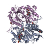 6kmmS S: Starting model for refinement |
|---|---|
| Similar structure data |
-Links
 PDBj
PDBj


- Assembly
| Deposited unit | 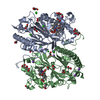
| ||||||||
|---|---|---|---|---|---|---|---|---|---|
| 1 |
| ||||||||
| Unit cell |
|
-Components
-Protein , 1 types, 2 molecules AB
| #1: Protein | Mass: 40235.324 Da / Num. of mol.: 2 Source method: isolated from a genetically manipulated source Source: (gene. exp.) Bacillus subtilis (bacteria) / Gene: efeB, GS595_16230 / Production host: Escherichia coli BL21(DE3) (bacteria)References: UniProt: A0A6G2J275, Oxidoreductases; Acting on a peroxide as acceptor; Peroxidases |
|---|
-Non-polymers , 9 types, 613 molecules 


| #2: Chemical | Heme B Mass: 616.487 Da / Num. of mol.: 2 / Source method: obtained synthetically / Formula: C34H32FeN4O4 Mass: 616.487 Da / Num. of mol.: 2 / Source method: obtained synthetically / Formula: C34H32FeN4O4#3: Chemical | Oxygen Mass: 31.999 Da / Num. of mol.: 2 / Source method: obtained synthetically / Formula: O2 Mass: 31.999 Da / Num. of mol.: 2 / Source method: obtained synthetically / Formula: O2#4: Chemical | ChemComp-EDO / Ethylene glycol Mass: 62.068 Da / Num. of mol.: 18 / Source method: obtained synthetically / Formula: C2H6O2 Mass: 62.068 Da / Num. of mol.: 18 / Source method: obtained synthetically / Formula: C2H6O2#5: Chemical | Citric acid Mass: 192.124 Da / Num. of mol.: 2 / Source method: obtained synthetically / Formula: C6H8O7 Mass: 192.124 Da / Num. of mol.: 2 / Source method: obtained synthetically / Formula: C6H8O7#6: Chemical | Diethylene glycol Mass: 106.120 Da / Num. of mol.: 3 / Source method: obtained synthetically / Formula: C4H10O3 Mass: 106.120 Da / Num. of mol.: 3 / Source method: obtained synthetically / Formula: C4H10O3#7: Chemical | ChemComp-NA /  Mass: 22.990 Da / Num. of mol.: 8 / Source method: obtained synthetically / Formula: Na Mass: 22.990 Da / Num. of mol.: 8 / Source method: obtained synthetically / Formula: Na#8: Chemical | ChemComp-CL / Chloride Mass: 35.453 Da / Num. of mol.: 9 / Source method: obtained synthetically / Formula: Cl Mass: 35.453 Da / Num. of mol.: 9 / Source method: obtained synthetically / Formula: Cl#9: Chemical | ChemComp-GOL / | Glycerol Mass: 92.094 Da / Num. of mol.: 1 / Source method: obtained synthetically / Formula: C3H8O3 Mass: 92.094 Da / Num. of mol.: 1 / Source method: obtained synthetically / Formula: C3H8O3#10: Water | ChemComp-HOH / | WaterMass: 18.015 Da / Num. of mol.: 568 / Source method: isolated from a natural source / Formula: H2O |
|---|
-Details
| Has ligand of interest | N |
|---|
-Experimental details
-Experiment
| Experiment | Method: X-RAY DIFFRACTION / Number of used crystals: 1 |
|---|
- Sample preparation
| Crystal | Density Matthews: 1.99 Å3/Da / Density % sol: 38.2 % |
|---|---|
| Crystal grow | Temperature: 293 K / Method: vapor diffusion, sitting drop / pH: 4 / Details: 0.1M Sodium citrate, 1M LiCl, 20% PEG 6000 |
-Data collection
| Diffraction | Mean temperature: 100 K / Serial crystal experiment: N |
|---|---|
| Diffraction source | Source: SYNCHROTRON / Site: ESRF  / Beamline: ID29 / Wavelength: 0.9677 Å / Beamline: ID29 / Wavelength: 0.9677 Å |
| Detector | Type: DECTRIS EIGER R 4M / Detector: PIXEL / Date: Nov 18, 2020 |
| Radiation | Protocol: SINGLE WAVELENGTH / Monochromatic (M) / Laue (L): M / Scattering type: x-ray |
| Radiation wavelength | Wavelength: 0.9677 Å / Relative weight: 1 |
| Reflection | Resolution: 1.9→44.344 Å / Num. obs: 52281 / % possible obs: 99.37 % / Redundancy: 6.8 % / CC1/2: 0.98 / Net I/σ(I): 8.8 |
| Reflection shell | Resolution: 1.9→1.96 Å / Num. unique obs: 7225 / CC1/2: 0.89 / % possible all: 5.8 |
- Processing
| Software |
| ||||||||||||||||||||||||||||||||||||||||||||||||||||||||||||||||||||||||||||||||||||||||||
|---|---|---|---|---|---|---|---|---|---|---|---|---|---|---|---|---|---|---|---|---|---|---|---|---|---|---|---|---|---|---|---|---|---|---|---|---|---|---|---|---|---|---|---|---|---|---|---|---|---|---|---|---|---|---|---|---|---|---|---|---|---|---|---|---|---|---|---|---|---|---|---|---|---|---|---|---|---|---|---|---|---|---|---|---|---|---|---|---|---|---|---|
| Refinement | Method to determine structure: MOLECULAR REPLACEMENT Starting model: 6KMM Resolution: 1.9→44.344 Å / SU ML: 0.22 / Cross valid method: THROUGHOUT / σ(F): 1.54 / Phase error: 27.39 / Stereochemistry target values: ML
| ||||||||||||||||||||||||||||||||||||||||||||||||||||||||||||||||||||||||||||||||||||||||||
| Solvent computation | Shrinkage radii: 0.9 Å / VDW probe radii: 1.11 Å / Solvent model: FLAT BULK SOLVENT MODEL | ||||||||||||||||||||||||||||||||||||||||||||||||||||||||||||||||||||||||||||||||||||||||||
| Displacement parameters | Biso max: 89.05 Å2 / Biso mean: 39.6611 Å2 / Biso min: 19.75 Å2 | ||||||||||||||||||||||||||||||||||||||||||||||||||||||||||||||||||||||||||||||||||||||||||
| Refinement step | Cycle: final / Resolution: 1.9→44.344 Å
| ||||||||||||||||||||||||||||||||||||||||||||||||||||||||||||||||||||||||||||||||||||||||||
| Refine LS restraints |
| ||||||||||||||||||||||||||||||||||||||||||||||||||||||||||||||||||||||||||||||||||||||||||
| LS refinement shell | Refine-ID: X-RAY DIFFRACTION / Rfactor Rfree error: 0
|
