 Movie
Movie Controller


+ Open data

- Basic information
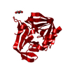
| Entry | Database: PDB / ID: 7e0w | ||||||
|---|---|---|---|---|---|---|---|
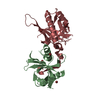
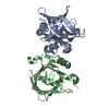
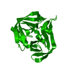
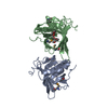
| Title | Crystal Structure of BCH domain from S. pombe | ||||||
 Components Components | Putative Rho GTPase-activating protein C1565.02c | ||||||
 Keywords Keywords | LIPID BINDING PROTEIN /  Intertwined / Anti-parallel / Dimeric structure Intertwined / Anti-parallel / Dimeric structure | ||||||
| Function / homology |  Function and homology information Function and homology information: / phospholipid transfer activity / intermembrane phospholipid transfer / GTPase activator activity / Golgi apparatus / signal transductionSimilarity search - Function | ||||||
| Biological species |  Schizosaccharomyces pombe 972h- (yeast) Schizosaccharomyces pombe 972h- (yeast) | ||||||
| Method | X-RAY DIFFRACTION / SYNCHROTRON / SAD / Resolution: 2.8 Å | ||||||
 Authors Authors | Chichili, V.P.R. / Jobichen, C. / Sivaraman, J. | ||||||
| Funding support |  Singapore, 1items Singapore, 1items
| ||||||
 Citation Citation | Journal: Proc.Natl.Acad.Sci.USA / Year: 2021 Title: A novel intertwined anti-parallel dimeric structure of scaffold BCH domain regulates RhoA and RhoGAP functions Authors: Chichili, V.P.R. / Chew, T.W. / Er, S.Y. / Chin, C.F. / Shankar, S. / Jobichen, C. / Pan, Q.C. / Zhou, Y.T. / Yeong, F.M. / Low, B.C. / Sivaraman, J. | ||||||
| History |
|
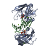
- Structure visualization
| Structure viewer | Molecule: MolmilJmol/JSmol |
|---|
- Downloads & links
-Download
| PDBx/mmCIF format | 7e0w.cif.gz | 133.3 KB | Display | PDBx/mmCIF format |
|---|---|---|---|---|
| PDB format | pdb7e0w.ent.gz | 104.8 KB | Display | PDB format |
| PDBx/mmJSON format | 7e0w.json.gz | Tree view | PDBx/mmJSON format | |
| Others |  Other downloads Other downloads |
-Validation report
| Arichive directory | https://data.pdbj.org/pub/pdb/validation_reports/e0/7e0wftp://data.pdbj.org/pub/pdb/validation_reports/e0/7e0w | HTTPS FTP |
|---|
-Related structure data
| Similar structure data |
|---|


-Links
 PDBj
PDBj
- Assembly
| Deposited unit | 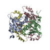
| ||||||||
|---|---|---|---|---|---|---|---|---|---|
| 1 | 
| ||||||||
| 2 | 
| ||||||||
| Unit cell |
|
-Components
| #1: Protein | Mass: 18716.518 Da / Num. of mol.: 4 Source method: isolated from a genetically manipulated source Source: (gene. exp.) Schizosaccharomyces pombe 972h- (yeast)Strain: 972 / ATCC 24843 / Gene: SPAC1565.02c / Production host:  Escherichia coli (E. coli) / References: UniProt: Q9P3B1 Escherichia coli (E. coli) / References: UniProt: Q9P3B1#2: Chemical | ChemComp-PG4 / Polyethylene glycol  Mass: 194.226 Da / Num. of mol.: 4 / Source method: obtained synthetically / Formula: C8H18O5 / Comment: precipitant*YM Mass: 194.226 Da / Num. of mol.: 4 / Source method: obtained synthetically / Formula: C8H18O5 / Comment: precipitant*YMHas ligand of interest | N | |
|---|
-Experimental details
-Experiment
| Experiment | Method: X-RAY DIFFRACTION / Number of used crystals: 1 |
|---|
- Sample preparation
| Crystal | Density Matthews: 5.67 Å3/Da / Density % sol: 78.31 % |
|---|---|
| Crystal grow | Temperature: 295 K / Method: vapor diffusion, hanging drop / Details: 0.1M Bis-Tris propane pH 7.0 and 2.1M NaCl. |
-Data collection
| Diffraction | Mean temperature: 100 K / Serial crystal experiment: N |
|---|---|
| Diffraction source | Source: SYNCHROTRON / Site: APS  / Beamline: 24-ID-C / Wavelength: 0.9789 Å / Beamline: 24-ID-C / Wavelength: 0.9789 Å |
| Detector | Type: DECTRIS PILATUS3 S 6M / Detector: PIXEL / Date: Jul 15, 2015 |
| Radiation | Protocol: SINGLE WAVELENGTH / Monochromatic (M) / Laue (L): M / Scattering type: x-ray |
| Radiation wavelength | Wavelength: 0.9789 Å / Relative weight: 1 |
| Reflection | Resolution: 2.8→50 Å / Num. obs: 36910 / % possible obs: 87.7 % / Redundancy: 5.5 % / Rsym value: 0.089 / Net I/σ(I): 12.6 |
| Reflection shell | Resolution: 2.8→2.93 Å / Num. unique obs: 2453 / Rsym value: 0.338 |
- Processing
| Software |
| |||||||||||||||||||||||||||||||||||||||||||||||||||||||||||||||||||||||||||||||||||||||||||||||||||||||||
|---|---|---|---|---|---|---|---|---|---|---|---|---|---|---|---|---|---|---|---|---|---|---|---|---|---|---|---|---|---|---|---|---|---|---|---|---|---|---|---|---|---|---|---|---|---|---|---|---|---|---|---|---|---|---|---|---|---|---|---|---|---|---|---|---|---|---|---|---|---|---|---|---|---|---|---|---|---|---|---|---|---|---|---|---|---|---|---|---|---|---|---|---|---|---|---|---|---|---|---|---|---|---|---|---|---|---|
| Refinement | Method to determine structure: SAD / Resolution: 2.8→49.736 Å / Cross valid method: FREE R-VALUE / σ(F): 146.76 / Phase error: 25.71
| |||||||||||||||||||||||||||||||||||||||||||||||||||||||||||||||||||||||||||||||||||||||||||||||||||||||||
| Solvent computation | Shrinkage radii: 0.9 Å / VDW probe radii: 1.11 Å | |||||||||||||||||||||||||||||||||||||||||||||||||||||||||||||||||||||||||||||||||||||||||||||||||||||||||
| Refinement step | Cycle: LAST / Resolution: 2.8→49.736 Å
| |||||||||||||||||||||||||||||||||||||||||||||||||||||||||||||||||||||||||||||||||||||||||||||||||||||||||
| Refine LS restraints |
| |||||||||||||||||||||||||||||||||||||||||||||||||||||||||||||||||||||||||||||||||||||||||||||||||||||||||
| LS refinement shell |
|
