 Movie
Movie Controller

[English] 日本語
 Yorodumi
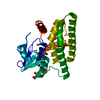
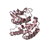
- PDB-7dw3: Crystal structure of a glutathione S-transferase mutant SbGSTU6(I... -
+ Open data

- Basic information
| Entry | Database: PDB / ID: 7dw3 | ||||||
|---|---|---|---|---|---|---|---|
| Title | Crystal structure of a glutathione S-transferase mutant SbGSTU6(I55T) from Salix babylonica | ||||||
 Components Components | Glutathione S-transferase | ||||||
 Keywords Keywords | TRANSFERASE / Enzyme / Complex / Mutant | ||||||
| Function / homology |  Function and homology information Function and homology informationresponse to chemical / glutathione transferase / glutathione transferase activity / glutathione metabolic process / cytosolSimilarity search - Function | ||||||
| Biological species |  Salix babylonica (weeping willow) Salix babylonica (weeping willow) | ||||||
| Method | X-RAY DIFFRACTION / SYNCHROTRON / MOLECULAR REPLACEMENT / Resolution: 1.93 Å | ||||||
 Authors Authors | Zhuge, X.L. / Yang, H.L. | ||||||
| Funding support |  China, 1items China, 1items
| ||||||
 Citation Citation | Journal: To Be Published Title: Crystal structure of a glutathione S-transferase SbGSTU6 from Salix babylonica in complex with glutathione Authors: Zhuge, X.L. / Yang, H.L. | ||||||
| History |
|
- Structure visualization
| Structure viewer | Molecule: MolmilJmol/JSmol |
|---|
- Downloads & links
-Download
| PDBx/mmCIF format | 7dw3.cif.gz | 60.5 KB | Display | PDBx/mmCIF format |
|---|---|---|---|---|
| PDB format | pdb7dw3.ent.gz | 41.3 KB | Display | PDB format |
| PDBx/mmJSON format | 7dw3.json.gz | Tree view | PDBx/mmJSON format | |
| Others |  Other downloads Other downloads |
-Validation report
| Arichive directory | https://data.pdbj.org/pub/pdb/validation_reports/dw/7dw3ftp://data.pdbj.org/pub/pdb/validation_reports/dw/7dw3 | HTTPS FTP |
|---|
-Related structure data
| Related structure data |  7dw1C 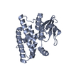 7dw2S S: Starting model for refinement C: citing same article ( |
|---|---|
| Similar structure data |


-Links
 PDBj
PDBj

- Assembly
| Deposited unit | 
| ||||||||||||
|---|---|---|---|---|---|---|---|---|---|---|---|---|---|
| 1 |
| ||||||||||||
| Unit cell |
| ||||||||||||
| Components on special symmetry positions |
|
-Components
| #1: Protein | Mass: 26342.508 Da / Num. of mol.: 1 / Mutation: I55T Source method: isolated from a genetically manipulated source Source: (gene. exp.) Salix babylonica (weeping willow) / Gene: GSTU6 / Production host:  Escherichia coli (E. coli) / References: UniProt: A0A4Y5R032 Escherichia coli (E. coli) / References: UniProt: A0A4Y5R032 |
|---|---|
| #2: Water | ChemComp-HOH / Water Mass: 18.015 Da / Num. of mol.: 109 / Source method: isolated from a natural source / Formula: H2O Mass: 18.015 Da / Num. of mol.: 109 / Source method: isolated from a natural source / Formula: H2O |
-Experimental details
-Experiment
| Experiment | Method: X-RAY DIFFRACTION / Number of used crystals: 1 |
|---|
- Sample preparation
| Crystal | Density Matthews: 3.19 Å3/Da / Density % sol: 61.49 % |
|---|---|
| Crystal grow | Temperature: 289.15 K / Method: vapor diffusion, hanging drop / pH: 7.4 Details: Magnesium acetate tetrahydrate, Sodium cacodylate trihydrate, PEG 8000 |
-Data collection
| Diffraction | Mean temperature: 100 K / Serial crystal experiment: N |
|---|---|
| Diffraction source | Source: SYNCHROTRON / Site: NFPSS / Beamline: BL19U1 / Wavelength: 0.987 Å |
| Detector | Type: DECTRIS PILATUS 6M / Detector: PIXEL / Date: Dec 1, 2019 |
| Radiation | Protocol: SINGLE WAVELENGTH / Monochromatic (M) / Laue (L): M / Scattering type: x-ray |
| Radiation wavelength | Wavelength: 0.987 Å / Relative weight: 1 |
| Reflection | Resolution: 1.93→34.51 Å / Num. obs: 25535 / % possible obs: 99.92 % / Redundancy: 1 % / Biso Wilson estimate: 32.59 Å2 / CC1/2: 1 / Net I/σ(I): 1.89 |
| Reflection shell | Resolution: 1.93→2.06 Å / Num. unique obs: 2513 / CC1/2: 1 |
- Processing
| Software |
| |||||||||||||||||||||||||||||||||||||||||||||||||||||||||||||||||||||||||||||||||||||||||||||||||||||||||
|---|---|---|---|---|---|---|---|---|---|---|---|---|---|---|---|---|---|---|---|---|---|---|---|---|---|---|---|---|---|---|---|---|---|---|---|---|---|---|---|---|---|---|---|---|---|---|---|---|---|---|---|---|---|---|---|---|---|---|---|---|---|---|---|---|---|---|---|---|---|---|---|---|---|---|---|---|---|---|---|---|---|---|---|---|---|---|---|---|---|---|---|---|---|---|---|---|---|---|---|---|---|---|---|---|---|---|
| Refinement | Method to determine structure: MOLECULAR REPLACEMENT Starting model: 7DW2 Resolution: 1.93→34.51 Å / SU ML: 0.2065 / Cross valid method: FREE R-VALUE / σ(F): 1.38 / Phase error: 25.9098 Stereochemistry target values: GeoStd + Monomer Library + CDL v1.2
| |||||||||||||||||||||||||||||||||||||||||||||||||||||||||||||||||||||||||||||||||||||||||||||||||||||||||
| Solvent computation | Shrinkage radii: 0.9 Å / VDW probe radii: 1.11 Å / Solvent model: FLAT BULK SOLVENT MODEL | |||||||||||||||||||||||||||||||||||||||||||||||||||||||||||||||||||||||||||||||||||||||||||||||||||||||||
| Displacement parameters | Biso mean: 37.18 Å2 | |||||||||||||||||||||||||||||||||||||||||||||||||||||||||||||||||||||||||||||||||||||||||||||||||||||||||
| Refinement step | Cycle: LAST / Resolution: 1.93→34.51 Å
| |||||||||||||||||||||||||||||||||||||||||||||||||||||||||||||||||||||||||||||||||||||||||||||||||||||||||
| Refine LS restraints |
| |||||||||||||||||||||||||||||||||||||||||||||||||||||||||||||||||||||||||||||||||||||||||||||||||||||||||
| LS refinement shell |
|
