 Movie
Movie Controller

[English] 日本語
 Yorodumi
- PDB-7ds7: The Crystal Structure of Leaf-branch compost cutinase from Biortus. -
+ Open data

- Basic information
| Entry | Database: PDB / ID: 7ds7 | ||||||
|---|---|---|---|---|---|---|---|
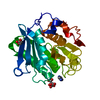
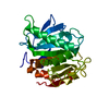
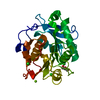
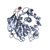
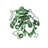
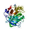
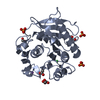
| Title | The Crystal Structure of Leaf-branch compost cutinase from Biortus. | ||||||
 Components Components | Leaf-branch compost cutinase | ||||||
 Keywords Keywords |  HYDROLASE / catalytic activity / hydrolase activity / acting on ester bonds HYDROLASE / catalytic activity / hydrolase activity / acting on ester bonds | ||||||
| Function / homology |  Function and homology information Function and homology informationpoly(ethylene terephthalate) hydrolase / acetylesterase activity / cutinase activity / cutinase / extracellular regionSimilarity search - Function | ||||||
| Biological species |  Unknown prokaryotic organism (environmental samples) Unknown prokaryotic organism (environmental samples) | ||||||
| Method | X-RAY DIFFRACTION / MOLECULAR REPLACEMENT / Resolution: 2.15 Å | ||||||
 Authors Authors | Wang, F. / Lv, Z. / Cheng, W. / Lin, D. / Chu, F. / Xu, X. / Tan, J. | ||||||
 Citation Citation | Journal: To Be Published Title: The Crystal Structure of Leaf-branch compost cutinase from Biortus. Authors: Wang, F. / Lv, Z. / Cheng, W. / Lin, D. / Chu, F. / Xu, X. / Tan, J. | ||||||
| History |
|
- Structure visualization
| Structure viewer | Molecule: MolmilJmol/JSmol |
|---|
- Downloads & links
-Download
| PDBx/mmCIF format | 7ds7.cif.gz | 68.6 KB | Display | PDBx/mmCIF format |
|---|---|---|---|---|
| PDB format | pdb7ds7.ent.gz | 45.8 KB | Display | PDB format |
| PDBx/mmJSON format | 7ds7.json.gz | Tree view | PDBx/mmJSON format | |
| Others |  Other downloads Other downloads |
-Validation report
| Arichive directory | https://data.pdbj.org/pub/pdb/validation_reports/ds/7ds7ftp://data.pdbj.org/pub/pdb/validation_reports/ds/7ds7 | HTTPS FTP |
|---|
-Related structure data
| Related structure data |  6thtS S: Starting model for refinement |
|---|---|
| Similar structure data |


-Links
 PDBj
PDBj


- Assembly
| Deposited unit | 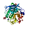
| ||||||||
|---|---|---|---|---|---|---|---|---|---|
| 1 |
| ||||||||
| Unit cell |
|
-Components
| #1: Protein | Mass: 29805.250 Da / Num. of mol.: 1 / Mutation: Y127G,S165A,D238C,F243I,S283C Source method: isolated from a genetically manipulated source Source: (gene. exp.) Unknown prokaryotic organism (environmental samples)Production host: Escherichia coli (E. coli)References: UniProt: G9BY57, cutinase, poly(ethylene terephthalate) hydrolase |
|---|---|
| #2: Chemical | ChemComp-IMD / Imidazole  Mass: 69.085 Da / Num. of mol.: 1 / Source method: obtained synthetically / Formula: C3H5N2 Mass: 69.085 Da / Num. of mol.: 1 / Source method: obtained synthetically / Formula: C3H5N2 |
| #3: Chemical | ChemComp-GOL / Glycerol  Mass: 92.094 Da / Num. of mol.: 1 / Source method: obtained synthetically / Formula: C3H8O3 Mass: 92.094 Da / Num. of mol.: 1 / Source method: obtained synthetically / Formula: C3H8O3 |
| #4: Chemical | ChemComp-CIT / Citric acid  Mass: 192.124 Da / Num. of mol.: 1 / Source method: obtained synthetically / Formula: C6H8O7 Mass: 192.124 Da / Num. of mol.: 1 / Source method: obtained synthetically / Formula: C6H8O7 |
| #5: Water | ChemComp-HOH / Water Mass: 18.015 Da / Num. of mol.: 114 / Source method: isolated from a natural source / Formula: H2O Mass: 18.015 Da / Num. of mol.: 114 / Source method: isolated from a natural source / Formula: H2O |
| Has ligand of interest | N |
-Experimental details
-Experiment
| Experiment | Method: X-RAY DIFFRACTION / Number of used crystals: 1 |
|---|
- Sample preparation
| Crystal | Density Matthews: 2.04 Å3/Da / Density % sol: 39.62 % |
|---|---|
| Crystal grow | Temperature: 293 K / Method: vapor diffusion, sitting drop / Details: 0.6M imidazole, 0.1M tri-sodium citrate PH7.8 |
-Data collection
| Diffraction | Mean temperature: 100 K / Serial crystal experiment: N |
|---|---|
| Diffraction source | Source: ROTATING ANODE / Type: RIGAKU FR-E SUPERBRIGHT / Wavelength: 1.54178 Å |
| Detector | Type: RIGAKU SATURN 944+ / Detector: CCD / Date: Nov 17, 2020 |
| Radiation | Protocol: SINGLE WAVELENGTH / Monochromatic (M) / Laue (L): M / Scattering type: x-ray |
| Radiation wavelength | Wavelength: 1.54178 Å / Relative weight: 1 |
| Reflection | Resolution: 2.15→47.38 Å / Num. obs: 13146 / % possible obs: 98.1 % / Redundancy: 11 % / Rmerge(I) obs: 0.101 / Net I/σ(I): 22.1 |
| Reflection shell | Resolution: 2.15→2.22 Å / Rmerge(I) obs: 0.262 / Num. unique obs: 1117 |
- Processing
| Software |
| ||||||||||||||||||||||||||||||||||||||||||||||||||||||||||||||||||||||||||||||||||||||||||||||||||||||||||||||||||||||||||||||||||||||||||||||||||||||
|---|---|---|---|---|---|---|---|---|---|---|---|---|---|---|---|---|---|---|---|---|---|---|---|---|---|---|---|---|---|---|---|---|---|---|---|---|---|---|---|---|---|---|---|---|---|---|---|---|---|---|---|---|---|---|---|---|---|---|---|---|---|---|---|---|---|---|---|---|---|---|---|---|---|---|---|---|---|---|---|---|---|---|---|---|---|---|---|---|---|---|---|---|---|---|---|---|---|---|---|---|---|---|---|---|---|---|---|---|---|---|---|---|---|---|---|---|---|---|---|---|---|---|---|---|---|---|---|---|---|---|---|---|---|---|---|---|---|---|---|---|---|---|---|---|---|---|---|---|---|---|---|
| Refinement | Method to determine structure: MOLECULAR REPLACEMENT Starting model: 6tht Resolution: 2.15→47.38 Å / Cor.coef. Fo:Fc: 0.937 / Cor.coef. Fo:Fc free: 0.904 / SU B: 4.606 / SU ML: 0.12 / Cross valid method: FREE R-VALUE / ESU R: 0.286 / ESU R Free: 0.206 Details: Hydrogens have been added in their riding positions
| ||||||||||||||||||||||||||||||||||||||||||||||||||||||||||||||||||||||||||||||||||||||||||||||||||||||||||||||||||||||||||||||||||||||||||||||||||||||
| Solvent computation | Ion probe radii: 0.8 Å / Shrinkage radii: 0.8 Å / VDW probe radii: 1.2 Å / Solvent model: MASK BULK SOLVENT | ||||||||||||||||||||||||||||||||||||||||||||||||||||||||||||||||||||||||||||||||||||||||||||||||||||||||||||||||||||||||||||||||||||||||||||||||||||||
| Displacement parameters | Biso mean: 9.937 Å2
| ||||||||||||||||||||||||||||||||||||||||||||||||||||||||||||||||||||||||||||||||||||||||||||||||||||||||||||||||||||||||||||||||||||||||||||||||||||||
| Refinement step | Cycle: LAST / Resolution: 2.15→47.38 Å
| ||||||||||||||||||||||||||||||||||||||||||||||||||||||||||||||||||||||||||||||||||||||||||||||||||||||||||||||||||||||||||||||||||||||||||||||||||||||
| Refine LS restraints |
| ||||||||||||||||||||||||||||||||||||||||||||||||||||||||||||||||||||||||||||||||||||||||||||||||||||||||||||||||||||||||||||||||||||||||||||||||||||||
| LS refinement shell |
|
