 Movie
Movie Controller

[English] 日本語
 Yorodumi
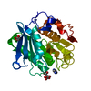
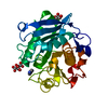
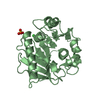
- PDB-6ths: High resolution crystal structure of Leaf-branch cutinase S165A v... -
+ Open data

- Basic information
| Entry | Database: PDB / ID: 6ths | ||||||
|---|---|---|---|---|---|---|---|
| Title | High resolution crystal structure of Leaf-branch cutinase S165A variant | ||||||
 Components Components | LCC | ||||||
 Keywords Keywords |  HYDROLASE / SERINE ESTERASE / CUTINASE HYDROLASE / SERINE ESTERASE / CUTINASE | ||||||
| Function / homology |  Function and homology information Function and homology informationpoly(ethylene terephthalate) hydrolase / acetylesterase activity / cutinase activity / cutinase / extracellular regionSimilarity search - Function | ||||||
| Biological species |  uncultured bacterium (environmental samples) uncultured bacterium (environmental samples) | ||||||
| Method | X-RAY DIFFRACTION / SYNCHROTRON / MOLECULAR REPLACEMENT / molecular replacement / Resolution: 1.1 Å | ||||||
 Authors Authors | Nomme, J. | ||||||
 Citation Citation | Journal: Nature / Year: 2020 Title: An engineered PET depolymerase to break down and recycle plastic bottles. Authors: Tournier, V. / Topham, C.M. / Gilles, A. / David, B. / Folgoas, C. / Moya-Leclair, E. / Kamionka, E. / Desrousseaux, M.L. / Texier, H. / Gavalda, S. / Cot, M. / Guemard, E. / Dalibey, M. / ...Authors: Tournier, V. / Topham, C.M. / Gilles, A. / David, B. / Folgoas, C. / Moya-Leclair, E. / Kamionka, E. / Desrousseaux, M.L. / Texier, H. / Gavalda, S. / Cot, M. / Guemard, E. / Dalibey, M. / Nomme, J. / Cioci, G. / Barbe, S. / Chateau, M. / Andre, I. / Duquesne, S. / Marty, A. | ||||||
| History |
|
- Structure visualization
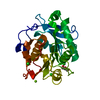
| Structure viewer | Molecule: MolmilJmol/JSmol |
|---|
- Downloads & links
-Download
| PDBx/mmCIF format | 6ths.cif.gz | 125.8 KB | Display | PDBx/mmCIF format |
|---|---|---|---|---|
| PDB format | pdb6ths.ent.gz | 95.3 KB | Display | PDB format |
| PDBx/mmJSON format | 6ths.json.gz | Tree view | PDBx/mmJSON format | |
| Others |  Other downloads Other downloads |
-Validation report
| Arichive directory | https://data.pdbj.org/pub/pdb/validation_reports/th/6thsftp://data.pdbj.org/pub/pdb/validation_reports/th/6ths | HTTPS FTP |
|---|
-Related structure data
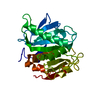
| Related structure data |  6thtC 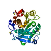 4eb0S S: Starting model for refinement C: citing same article ( |
|---|---|
| Similar structure data |


-Links
 PDBj
PDBj
- Assembly
| Deposited unit | 
| ||||||||
|---|---|---|---|---|---|---|---|---|---|
| 1 |
| ||||||||
| Unit cell |
|
-Components
| #1: Protein | Mass: 27783.992 Da / Num. of mol.: 1 / Mutation: S165A Source method: isolated from a genetically manipulated source Source: (gene. exp.) uncultured bacterium (environmental samples)Plasmid: pET26b / Production host: Escherichia coli BL21(DE3) (bacteria) / References: UniProt: G9BY57, cutinase |
|---|---|
| #2: Chemical | ChemComp-DIO / 1,4-Dioxane  Mass: 88.105 Da / Num. of mol.: 1 / Source method: obtained synthetically / Formula: C4H8O2 Mass: 88.105 Da / Num. of mol.: 1 / Source method: obtained synthetically / Formula: C4H8O2 |
| #3: Water | ChemComp-HOH / Water Mass: 18.015 Da / Num. of mol.: 290 / Source method: isolated from a natural source / Formula: H2O Mass: 18.015 Da / Num. of mol.: 290 / Source method: isolated from a natural source / Formula: H2O |
| Has ligand of interest | N |
-Experimental details
-Experiment
| Experiment | Method: X-RAY DIFFRACTION / Number of used crystals: 1 |
|---|
- Sample preparation
| Crystal | Density Matthews: 2.2 Å3/Da / Density % sol: 44.15 % |
|---|---|
| Crystal grow | Temperature: 285 K / Method: vapor diffusion / pH: 9 / Details: 0.1 M Bicine pH 9.0, 2% Dioxane, 12% PEG 20000 |
-Data collection
| Diffraction | Mean temperature: 100 K / Serial crystal experiment: N | ||||||||||||||||||||||||||||||||||||||||||||||||||||||||||||||||||||||||||||||||
|---|---|---|---|---|---|---|---|---|---|---|---|---|---|---|---|---|---|---|---|---|---|---|---|---|---|---|---|---|---|---|---|---|---|---|---|---|---|---|---|---|---|---|---|---|---|---|---|---|---|---|---|---|---|---|---|---|---|---|---|---|---|---|---|---|---|---|---|---|---|---|---|---|---|---|---|---|---|---|---|---|---|
| Diffraction source | Source: SYNCHROTRON / Site: ALBA  / Beamline: XALOC / Wavelength: 0.97907 Å / Beamline: XALOC / Wavelength: 0.97907 Å | ||||||||||||||||||||||||||||||||||||||||||||||||||||||||||||||||||||||||||||||||
| Detector | Type: DECTRIS PILATUS 6M / Detector: PIXEL / Date: Jun 2, 2018 | ||||||||||||||||||||||||||||||||||||||||||||||||||||||||||||||||||||||||||||||||
| Radiation | Protocol: SINGLE WAVELENGTH / Monochromatic (M) / Laue (L): M / Scattering type: x-ray | ||||||||||||||||||||||||||||||||||||||||||||||||||||||||||||||||||||||||||||||||
| Radiation wavelength | Wavelength: 0.97907 Å / Relative weight: 1 | ||||||||||||||||||||||||||||||||||||||||||||||||||||||||||||||||||||||||||||||||
| Reflection | Resolution: 1.1→47.51 Å / Num. obs: 98700 / % possible obs: 99.6 % / Redundancy: 12.711 % / Biso Wilson estimate: 15.012 Å2 / CC1/2: 0.999 / Rmerge(I) obs: 0.067 / Rrim(I) all: 0.07 / Χ2: 1.152 / Net I/σ(I): 18.06 | ||||||||||||||||||||||||||||||||||||||||||||||||||||||||||||||||||||||||||||||||
| Reflection shell | Diffraction-ID: 1
|
-Phasing
| Phasing | Method: molecular replacement |
|---|
- Processing
| Software |
| |||||||||||||||||||||||||||||||||||||||||||||||||||||||||||||||||||||||||||
|---|---|---|---|---|---|---|---|---|---|---|---|---|---|---|---|---|---|---|---|---|---|---|---|---|---|---|---|---|---|---|---|---|---|---|---|---|---|---|---|---|---|---|---|---|---|---|---|---|---|---|---|---|---|---|---|---|---|---|---|---|---|---|---|---|---|---|---|---|---|---|---|---|---|---|---|---|
| Refinement | Method to determine structure: MOLECULAR REPLACEMENT Starting model: 4EB0 Resolution: 1.1→47.51 Å / Cor.coef. Fo:Fc: 0.985 / Cor.coef. Fo:Fc free: 0.978 / SU B: 2.066 / SU ML: 0.039 / Cross valid method: THROUGHOUT / σ(F): 0 / ESU R: 0.03 / ESU R Free: 0.032 Details: HYDROGENS HAVE BEEN ADDED IN THE RIDING POSITIONS U VALUES : REFINED INDIVIDUALLY
| |||||||||||||||||||||||||||||||||||||||||||||||||||||||||||||||||||||||||||
| Solvent computation | Ion probe radii: 0.8 Å / Shrinkage radii: 0.8 Å / VDW probe radii: 1.2 Å | |||||||||||||||||||||||||||||||||||||||||||||||||||||||||||||||||||||||||||
| Displacement parameters | Biso max: 70.19 Å2 / Biso mean: 15.091 Å2 / Biso min: 9 Å2
| |||||||||||||||||||||||||||||||||||||||||||||||||||||||||||||||||||||||||||
| Refinement step | Cycle: final / Resolution: 1.1→47.51 Å
| |||||||||||||||||||||||||||||||||||||||||||||||||||||||||||||||||||||||||||
| Refine LS restraints |
| |||||||||||||||||||||||||||||||||||||||||||||||||||||||||||||||||||||||||||
| LS refinement shell | Resolution: 1.1→1.127 Å / Rfactor Rfree error: 0
|
