 Movie
Movie Controller

[English] 日本語
 Yorodumi
- PDB-7dnr: Crystal Structure of Zn-bound SIS Domain of Glucosamine-6-P Synth... -
+ Open data

- Basic information
| Entry | Database: PDB / ID: 7dnr | ||||||
|---|---|---|---|---|---|---|---|
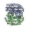
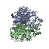
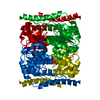
| Title | Crystal Structure of Zn-bound SIS Domain of Glucosamine-6-P Synthase from E. coli | ||||||
 Components Components | Glutamine--fructose-6-phosphate aminotransferase [isomerizing] | ||||||
 Keywords Keywords |  ISOMERASE / Metal binding / Zinc ion ISOMERASE / Metal binding / Zinc ion | ||||||
| Function / homology |  Function and homology informationglutamine-fructose-6-phosphate transaminase (isomerizing) / glutamine-fructose-6-phosphate transaminase (isomerizing) activity / UDP-N-acetylglucosamine metabolic process / UDP-N-acetylglucosamine biosynthetic process / carbohydrate derivative binding / fructose 6-phosphate metabolic process / protein N-linked glycosylation / glutamine metabolic process / carbohydrate metabolic process / cytosol Function and homology informationglutamine-fructose-6-phosphate transaminase (isomerizing) / glutamine-fructose-6-phosphate transaminase (isomerizing) activity / UDP-N-acetylglucosamine metabolic process / UDP-N-acetylglucosamine biosynthetic process / carbohydrate derivative binding / fructose 6-phosphate metabolic process / protein N-linked glycosylation / glutamine metabolic process / carbohydrate metabolic process / cytosolSimilarity search - Function | ||||||
| Biological species |  Escherichia coli (E. coli) Escherichia coli (E. coli) | ||||||
| Method | X-RAY DIFFRACTION / SYNCHROTRON / MOLECULAR REPLACEMENT / molecular replacement / Resolution: 1.7 Å | ||||||
 Authors Authors | Gao, C. / Xiao, J. | ||||||
 Citation Citation | Journal: To Be Published Title: Discovery of Metal-binding Proteins by Thermal Proteome Profiling Authors: Zeng, X. / Wei, T. / Liu, Y. / Wang, X. / Feng, T. / Cheng, Y. / Qin, W. / Gao, C. / Xiao, J. / Wang, C. | ||||||
| History |
|
- Structure visualization
| Structure viewer | Molecule: MolmilJmol/JSmol |
|---|
- Downloads & links
-Download
| PDBx/mmCIF format | 7dnr.cif.gz | 300.5 KB | Display | PDBx/mmCIF format |
|---|---|---|---|---|
| PDB format | pdb7dnr.ent.gz | 242.8 KB | Display | PDB format |
| PDBx/mmJSON format | 7dnr.json.gz | Tree view | PDBx/mmJSON format | |
| Others |  Other downloads Other downloads |
-Validation report
| Arichive directory | https://data.pdbj.org/pub/pdb/validation_reports/dn/7dnrftp://data.pdbj.org/pub/pdb/validation_reports/dn/7dnr | HTTPS FTP |
|---|
-Related structure data
| Related structure data | 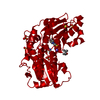 1moqS S: Starting model for refinement |
|---|---|
| Similar structure data |

-Links
 PDBj
PDBj


- Assembly
| Deposited unit | 
| ||||||||
|---|---|---|---|---|---|---|---|---|---|
| 1 |
| ||||||||
| Unit cell |
|
-Components
| #1: Protein | Mass: 40357.004 Da / Num. of mol.: 2 Source method: isolated from a genetically manipulated source Source: (gene. exp.) Escherichia coli (strain K12) (bacteria)Gene: glmS, b3729, JW3707 / Production host: Escherichia coli (E. coli)References: UniProt: P17169, glutamine-fructose-6-phosphate transaminase (isomerizing)#2: Chemical |   Mass: 65.409 Da / Num. of mol.: 3 / Source method: obtained synthetically / Formula: Zn / Feature type: SUBJECT OF INVESTIGATION Mass: 65.409 Da / Num. of mol.: 3 / Source method: obtained synthetically / Formula: Zn / Feature type: SUBJECT OF INVESTIGATION#3: Water | ChemComp-HOH / | Water Mass: 18.015 Da / Num. of mol.: 489 / Source method: isolated from a natural source / Formula: H2O Mass: 18.015 Da / Num. of mol.: 489 / Source method: isolated from a natural source / Formula: H2OHas ligand of interest | Y | |
|---|
-Experimental details
-Experiment
| Experiment | Method: X-RAY DIFFRACTION / Number of used crystals: 1 |
|---|
- Sample preparation
| Crystal | Density Matthews: 2.09 Å3/Da / Density % sol: 41.2 % |
|---|---|
| Crystal grow | Temperature: 291.15 K / Method: vapor diffusion, sitting drop / pH: 5.5 / Details: PEG 5000 MME, sodium acetate, zinc cloride |
-Data collection
| Diffraction | Mean temperature: 100 K / Serial crystal experiment: N | |||||||||||||||||||||||||||||||||||||||||||||||||||||||||||||||||||||||||||||||||||||||||||||||||||||||||||||||||||||||||||||||||||||||||||||||||||||||||||||||||||||||||||||||||||||||||||||
|---|---|---|---|---|---|---|---|---|---|---|---|---|---|---|---|---|---|---|---|---|---|---|---|---|---|---|---|---|---|---|---|---|---|---|---|---|---|---|---|---|---|---|---|---|---|---|---|---|---|---|---|---|---|---|---|---|---|---|---|---|---|---|---|---|---|---|---|---|---|---|---|---|---|---|---|---|---|---|---|---|---|---|---|---|---|---|---|---|---|---|---|---|---|---|---|---|---|---|---|---|---|---|---|---|---|---|---|---|---|---|---|---|---|---|---|---|---|---|---|---|---|---|---|---|---|---|---|---|---|---|---|---|---|---|---|---|---|---|---|---|---|---|---|---|---|---|---|---|---|---|---|---|---|---|---|---|---|---|---|---|---|---|---|---|---|---|---|---|---|---|---|---|---|---|---|---|---|---|---|---|---|---|---|---|---|---|---|---|---|---|
| Diffraction source | Source: SYNCHROTRON / Site: SSRF  / Beamline: BL17U1 / Wavelength: 0.9792 Å / Beamline: BL17U1 / Wavelength: 0.9792 Å | |||||||||||||||||||||||||||||||||||||||||||||||||||||||||||||||||||||||||||||||||||||||||||||||||||||||||||||||||||||||||||||||||||||||||||||||||||||||||||||||||||||||||||||||||||||||||||||
| Detector | Type: DECTRIS EIGER X 16M / Detector: PIXEL / Date: Dec 28, 2019 | |||||||||||||||||||||||||||||||||||||||||||||||||||||||||||||||||||||||||||||||||||||||||||||||||||||||||||||||||||||||||||||||||||||||||||||||||||||||||||||||||||||||||||||||||||||||||||||
| Radiation | Protocol: SINGLE WAVELENGTH / Monochromatic (M) / Laue (L): M / Scattering type: x-ray | |||||||||||||||||||||||||||||||||||||||||||||||||||||||||||||||||||||||||||||||||||||||||||||||||||||||||||||||||||||||||||||||||||||||||||||||||||||||||||||||||||||||||||||||||||||||||||||
| Radiation wavelength | Wavelength: 0.9792 Å / Relative weight: 1 | |||||||||||||||||||||||||||||||||||||||||||||||||||||||||||||||||||||||||||||||||||||||||||||||||||||||||||||||||||||||||||||||||||||||||||||||||||||||||||||||||||||||||||||||||||||||||||||
| Reflection | Resolution: 1.6→50 Å / Num. obs: 85139 / % possible obs: 100 % / Redundancy: 6.8 % / Rmerge(I) obs: 0.046 / Rpim(I) all: 0.019 / Rrim(I) all: 0.05 / Χ2: 0.609 / Net I/σ(I): 7.5 / Num. measured all: 577746 | |||||||||||||||||||||||||||||||||||||||||||||||||||||||||||||||||||||||||||||||||||||||||||||||||||||||||||||||||||||||||||||||||||||||||||||||||||||||||||||||||||||||||||||||||||||||||||||
| Reflection shell | Diffraction-ID: 1
|
-Phasing
| Phasing | Method: molecular replacement | ||||||
|---|---|---|---|---|---|---|---|
| Phasing MR |
|
- Processing
| Software |
| ||||||||||||||||||||||||||||||||||||||||||||||||||||||||||||||||||||||||||||||
|---|---|---|---|---|---|---|---|---|---|---|---|---|---|---|---|---|---|---|---|---|---|---|---|---|---|---|---|---|---|---|---|---|---|---|---|---|---|---|---|---|---|---|---|---|---|---|---|---|---|---|---|---|---|---|---|---|---|---|---|---|---|---|---|---|---|---|---|---|---|---|---|---|---|---|---|---|---|---|---|
| Refinement | Method to determine structure: MOLECULAR REPLACEMENT Starting model: 1MOQ Resolution: 1.7→23.52 Å / SU ML: 0.21 / Cross valid method: THROUGHOUT / σ(F): 1.38 / Phase error: 25.95 / Stereochemistry target values: ML
| ||||||||||||||||||||||||||||||||||||||||||||||||||||||||||||||||||||||||||||||
| Solvent computation | Shrinkage radii: 0.9 Å / VDW probe radii: 1.11 Å / Solvent model: FLAT BULK SOLVENT MODEL | ||||||||||||||||||||||||||||||||||||||||||||||||||||||||||||||||||||||||||||||
| Displacement parameters | Biso max: 89.47 Å2 / Biso mean: 27.8161 Å2 / Biso min: 11.91 Å2 | ||||||||||||||||||||||||||||||||||||||||||||||||||||||||||||||||||||||||||||||
| Refinement step | Cycle: final / Resolution: 1.7→23.52 Å
| ||||||||||||||||||||||||||||||||||||||||||||||||||||||||||||||||||||||||||||||
| LS refinement shell | Refine-ID: X-RAY DIFFRACTION / Rfactor Rfree error: 0 / Total num. of bins used: 12 / % reflection obs: 100 %
|
