 Movie
Movie Controller

[English] 日本語
 Yorodumi
- PDB-3knz: Crystal structure of Putative sugar binding protein (NP_459565.1)... -
+ Open data

- Basic information
| Entry | Database: PDB / ID: 3knz | ||||||
|---|---|---|---|---|---|---|---|
| Title | Crystal structure of Putative sugar binding protein (NP_459565.1) from Salmonella typhimurium LT2 at 2.50 A resolution | ||||||
 Components Components | Putative sugar binding protein | ||||||
 Keywords Keywords | SUGAR BINDING PROTEIN / Putative sugar binding protein /  Structural Genomics / Joint Center for Structural Genomics / JCSG / Protein Structure Initiative / PSI-2 Structural Genomics / Joint Center for Structural Genomics / JCSG / Protein Structure Initiative / PSI-2 | ||||||
| Function / homology |  Function and homology informationglutamine-fructose-6-phosphate transaminase (isomerizing) activity / UDP-N-acetylglucosamine metabolic process / carbohydrate derivative binding / fructose 6-phosphate metabolic process / protein N-linked glycosylation Function and homology informationglutamine-fructose-6-phosphate transaminase (isomerizing) activity / UDP-N-acetylglucosamine metabolic process / carbohydrate derivative binding / fructose 6-phosphate metabolic process / protein N-linked glycosylationSimilarity search - Function | ||||||
| Biological species |  Salmonella enterica subsp. enterica serovar Typhimurium (bacteria) Salmonella enterica subsp. enterica serovar Typhimurium (bacteria) | ||||||
| Method | X-RAY DIFFRACTION / SYNCHROTRON / MAD / Resolution: 2.5 Å | ||||||
 Authors Authors | Joint Center for Structural Genomics (JCSG) | ||||||
 Citation Citation | Journal: To be published Title: Crystal structure of Putative sugar binding protein (NP_459565.1) from Salmonella typhimurium LT2 at 2.50 A resolution Authors: Joint Center for Structural Genomics (JCSG) | ||||||
| History |
|
- Structure visualization
| Structure viewer | Molecule: MolmilJmol/JSmol |
|---|
- Downloads & links
-Download
| PDBx/mmCIF format | 3knz.cif.gz | 375.9 KB | Display | PDBx/mmCIF format |
|---|---|---|---|---|
| PDB format | pdb3knz.ent.gz | 319.6 KB | Display | PDB format |
| PDBx/mmJSON format | 3knz.json.gz | Tree view | PDBx/mmJSON format | |
| Others |  Other downloads Other downloads |
-Validation report
| Arichive directory | https://data.pdbj.org/pub/pdb/validation_reports/kn/3knzftp://data.pdbj.org/pub/pdb/validation_reports/kn/3knz | HTTPS FTP |
|---|
-Related structure data
| Similar structure data | |
|---|---|
| Other databases |


-Links
 PDBj
PDBj


- Assembly
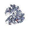
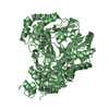
| Deposited unit | 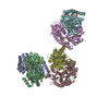
| |||||||||||||||||||||||||||||||||||||||||||||||||||||||||||||||
|---|---|---|---|---|---|---|---|---|---|---|---|---|---|---|---|---|---|---|---|---|---|---|---|---|---|---|---|---|---|---|---|---|---|---|---|---|---|---|---|---|---|---|---|---|---|---|---|---|---|---|---|---|---|---|---|---|---|---|---|---|---|---|---|---|
| 1 | 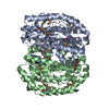
| |||||||||||||||||||||||||||||||||||||||||||||||||||||||||||||||
| 2 | 
| |||||||||||||||||||||||||||||||||||||||||||||||||||||||||||||||
| 3 | 
| |||||||||||||||||||||||||||||||||||||||||||||||||||||||||||||||
| Unit cell |
| |||||||||||||||||||||||||||||||||||||||||||||||||||||||||||||||
| Noncrystallographic symmetry (NCS) | NCS domain:
NCS domain segments:
| |||||||||||||||||||||||||||||||||||||||||||||||||||||||||||||||
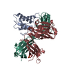
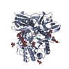
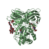
| Details | CRYSTAL PACKING ANALYSIS AND SIZE EXCLUSION CHROMATOGRAPHY COMBINED WITH STATIC LIGHT SCATTERING SUPPORT THE ASSIGNMENT OF A DIMER AS THE SIGNIFICANT OLIGOMERIC FORM IN SOLUTION. |
-Components
| #1: Protein | Mass: 41157.023 Da / Num. of mol.: 6 Source method: isolated from a genetically manipulated source Source: (gene. exp.) Salmonella enterica subsp. enterica serovar Typhimurium (bacteria)Gene: STM0573 / Plasmid: SpeedET / Production host: Escherichia Coli (E. coli) / Strain (production host): HK100 / References: UniProt: Q8ZR49#2: Chemical | 2-Ethoxyethanol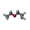  Mass: 90.121 Da / Num. of mol.: 2 / Source method: obtained synthetically / Formula: C4H10O2 Mass: 90.121 Da / Num. of mol.: 2 / Source method: obtained synthetically / Formula: C4H10O2#3: Chemical | Imidazole  Mass: 69.085 Da / Num. of mol.: 2 / Source method: obtained synthetically / Formula: C3H5N2 Mass: 69.085 Da / Num. of mol.: 2 / Source method: obtained synthetically / Formula: C3H5N2#4: Water | ChemComp-HOH / | Water Mass: 18.015 Da / Num. of mol.: 431 / Source method: isolated from a natural source / Formula: H2O Mass: 18.015 Da / Num. of mol.: 431 / Source method: isolated from a natural source / Formula: H2OSequence details | THIS CONSTRUCT WAS EXPRESSED WITH A PURIFICATI | |
|---|
-Experimental details
-Experiment
| Experiment | Method: X-RAY DIFFRACTION / Number of used crystals: 1 |
|---|
- Sample preparation
| Crystal | Density Matthews: 3.3 Å3/Da / Density % sol: 62.67 % |
|---|---|
| Crystal grow | Temperature: 277 K / Method: vapor diffusion, sitting drop / pH: 7.5 Details: 37.1000% 2-ethoxyethanol, 0.0500M calcium acetate, 0.1M Imidazole pH 7.5, NANODROP, VAPOR DIFFUSION, SITTING DROP, temperature 277K |
-Data collection
| Diffraction | Mean temperature: 100 K | ||||||||||||||||||||||||||||||||||||||||||||||||||||||||||||||||||||||||||||||||||||||||||||||||||||||||||||||||||||||||||||||||||||||||||||||||||||||||||||||||||||||||
|---|---|---|---|---|---|---|---|---|---|---|---|---|---|---|---|---|---|---|---|---|---|---|---|---|---|---|---|---|---|---|---|---|---|---|---|---|---|---|---|---|---|---|---|---|---|---|---|---|---|---|---|---|---|---|---|---|---|---|---|---|---|---|---|---|---|---|---|---|---|---|---|---|---|---|---|---|---|---|---|---|---|---|---|---|---|---|---|---|---|---|---|---|---|---|---|---|---|---|---|---|---|---|---|---|---|---|---|---|---|---|---|---|---|---|---|---|---|---|---|---|---|---|---|---|---|---|---|---|---|---|---|---|---|---|---|---|---|---|---|---|---|---|---|---|---|---|---|---|---|---|---|---|---|---|---|---|---|---|---|---|---|---|---|---|---|---|---|---|---|
| Diffraction source | Source: SYNCHROTRON / Site: SSRL  / Beamline: BL9-2 / Wavelength: 0.91837,0.97934,0.97922 / Beamline: BL9-2 / Wavelength: 0.91837,0.97934,0.97922 | ||||||||||||||||||||||||||||||||||||||||||||||||||||||||||||||||||||||||||||||||||||||||||||||||||||||||||||||||||||||||||||||||||||||||||||||||||||||||||||||||||||||||
| Detector | Type: MARMOSAIC 325 mm CCD / Detector: CCD / Date: Mar 20, 2009 / Details: Flat collimating mirror, toroid focusing mirror | ||||||||||||||||||||||||||||||||||||||||||||||||||||||||||||||||||||||||||||||||||||||||||||||||||||||||||||||||||||||||||||||||||||||||||||||||||||||||||||||||||||||||
| Radiation | Monochromator: Double crystal monochromator / Protocol: MAD / Monochromatic (M) / Laue (L): M / Scattering type: x-ray | ||||||||||||||||||||||||||||||||||||||||||||||||||||||||||||||||||||||||||||||||||||||||||||||||||||||||||||||||||||||||||||||||||||||||||||||||||||||||||||||||||||||||
| Radiation wavelength |
| ||||||||||||||||||||||||||||||||||||||||||||||||||||||||||||||||||||||||||||||||||||||||||||||||||||||||||||||||||||||||||||||||||||||||||||||||||||||||||||||||||||||||
| Reflection | Resolution: 2.5→29.907 Å / Num. obs: 113412 / % possible obs: 99.9 % / Redundancy: 3.8 % / Biso Wilson estimate: 48.836 Å2 / Rmerge(I) obs: 0.123 / Rsym value: 0.123 / Net I/σ(I): 9.6 | ||||||||||||||||||||||||||||||||||||||||||||||||||||||||||||||||||||||||||||||||||||||||||||||||||||||||||||||||||||||||||||||||||||||||||||||||||||||||||||||||||||||||
| Reflection shell | Diffraction-ID: 1
|
-Phasing
| Phasing | Method: MAD |
|---|
- Processing
| Software |
| |||||||||||||||||||||||||||||||||||||||||||||||||||||||||||||||||||||||||||||||||||||||||||||||||||||||||||||||||||||||||||||||||||||||||||||||||||||||||||||||||||||||||||||||
|---|---|---|---|---|---|---|---|---|---|---|---|---|---|---|---|---|---|---|---|---|---|---|---|---|---|---|---|---|---|---|---|---|---|---|---|---|---|---|---|---|---|---|---|---|---|---|---|---|---|---|---|---|---|---|---|---|---|---|---|---|---|---|---|---|---|---|---|---|---|---|---|---|---|---|---|---|---|---|---|---|---|---|---|---|---|---|---|---|---|---|---|---|---|---|---|---|---|---|---|---|---|---|---|---|---|---|---|---|---|---|---|---|---|---|---|---|---|---|---|---|---|---|---|---|---|---|---|---|---|---|---|---|---|---|---|---|---|---|---|---|---|---|---|---|---|---|---|---|---|---|---|---|---|---|---|---|---|---|---|---|---|---|---|---|---|---|---|---|---|---|---|---|---|---|---|---|
| Refinement | Method to determine structure: MAD / Resolution: 2.5→29.907 Å / Cor.coef. Fo:Fc: 0.943 / Cor.coef. Fo:Fc free: 0.929 / Occupancy max: 1 / Occupancy min: 0.5 / SU B: 17.422 / SU ML: 0.167 / TLS residual ADP flag: LIKELY RESIDUAL / Cross valid method: THROUGHOUT / σ(F): 0 / ESU R: 0.297 / ESU R Free: 0.222 Stereochemistry target values: MAXIMUM LIKELIHOOD WITH PHASES Details: 1.HYDROGENS HAVE BEEN ADDED IN THE RIDING POSITIONS. 2.ATOM RECORDS CONTAIN RESIDUAL B FACTORS ONLY. 3.A MET-INHIBITION PROTOCOL WAS USED FOR SELENOMETHIONINE INCORPORATION DURING PROTEIN ...Details: 1.HYDROGENS HAVE BEEN ADDED IN THE RIDING POSITIONS. 2.ATOM RECORDS CONTAIN RESIDUAL B FACTORS ONLY. 3.A MET-INHIBITION PROTOCOL WAS USED FOR SELENOMETHIONINE INCORPORATION DURING PROTEIN EXPRESSION. THE OCCUPANCY OF THE SE ATOMS IN THE MSE RESIDUES WAS REDUCED TO 0.75 FOR THE REDUCED SCATTERING POWER DUE TO PARTIAL S-MET INCORPORATION. 4.2-ETHOXYETHANOL (ETX) AND IMIDAZOLE (IMD) FROM THE CRYSTALLIZATION SOLUTION HAVE BEEN MODELED IN THE SOLVENT STRUCTURE.
| |||||||||||||||||||||||||||||||||||||||||||||||||||||||||||||||||||||||||||||||||||||||||||||||||||||||||||||||||||||||||||||||||||||||||||||||||||||||||||||||||||||||||||||||
| Solvent computation | Ion probe radii: 0.8 Å / Shrinkage radii: 0.8 Å / VDW probe radii: 1.2 Å / Solvent model: MASK | |||||||||||||||||||||||||||||||||||||||||||||||||||||||||||||||||||||||||||||||||||||||||||||||||||||||||||||||||||||||||||||||||||||||||||||||||||||||||||||||||||||||||||||||
| Displacement parameters | Biso max: 67.18 Å2 / Biso mean: 27.788 Å2 / Biso min: 3.85 Å2
| |||||||||||||||||||||||||||||||||||||||||||||||||||||||||||||||||||||||||||||||||||||||||||||||||||||||||||||||||||||||||||||||||||||||||||||||||||||||||||||||||||||||||||||||
| Refinement step | Cycle: LAST / Resolution: 2.5→29.907 Å
| |||||||||||||||||||||||||||||||||||||||||||||||||||||||||||||||||||||||||||||||||||||||||||||||||||||||||||||||||||||||||||||||||||||||||||||||||||||||||||||||||||||||||||||||
| Refine LS restraints |
| |||||||||||||||||||||||||||||||||||||||||||||||||||||||||||||||||||||||||||||||||||||||||||||||||||||||||||||||||||||||||||||||||||||||||||||||||||||||||||||||||||||||||||||||
| Refine LS restraints NCS | Dom-ID: 1 / Ens-ID: 1 / Number: 3917 / Refine-ID: X-RAY DIFFRACTION
| |||||||||||||||||||||||||||||||||||||||||||||||||||||||||||||||||||||||||||||||||||||||||||||||||||||||||||||||||||||||||||||||||||||||||||||||||||||||||||||||||||||||||||||||
| LS refinement shell | Resolution: 2.5→2.564 Å / Total num. of bins used: 20
| |||||||||||||||||||||||||||||||||||||||||||||||||||||||||||||||||||||||||||||||||||||||||||||||||||||||||||||||||||||||||||||||||||||||||||||||||||||||||||||||||||||||||||||||
| Refinement TLS params. | Method: refined / Refine-ID: X-RAY DIFFRACTION
| |||||||||||||||||||||||||||||||||||||||||||||||||||||||||||||||||||||||||||||||||||||||||||||||||||||||||||||||||||||||||||||||||||||||||||||||||||||||||||||||||||||||||||||||
| Refinement TLS group |
|
