 Movie
Movie Controller


+ Open data

- Basic information
| Entry | Database: PDB / ID: 7dk8 | ||||||
|---|---|---|---|---|---|---|---|
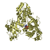
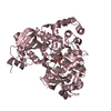
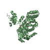
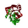
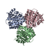
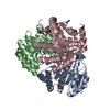
| Title | Crystal structure of OsGH3-8 with AMP | ||||||
 Components Components | Probable indole-3-acetic acid-amido synthetase GH3.8 | ||||||
 Keywords Keywords |  PLANT PROTEIN / acyl acid amido synthetases / auxin / active site PLANT PROTEIN / acyl acid amido synthetases / auxin / active site | ||||||
| Function / homology |  Function and homology information Function and homology informationindole-3-acetic acid amido synthetase activity / regulation of defense response to bacterium / : / Ligases; Forming carbon-nitrogen bonds; Acid-amino-acid ligases (peptide synthases) / pollen development / AMP binding / defense responseSimilarity search - Function | ||||||
| Biological species |  Oryza sativa subsp. indica (long-grained rice) Oryza sativa subsp. indica (long-grained rice) | ||||||
| Method | X-RAY DIFFRACTION / SYNCHROTRON / MOLECULAR REPLACEMENT / Resolution: 1.99 Å | ||||||
 Authors Authors | Zhang, Y.K. / Xu, G.L. / Ming, Z.H. | ||||||
| Funding support |  China, 1items China, 1items
| ||||||
 Citation Citation | Journal: Biochem.Biophys.Res.Commun. / Year: 2021 Title: Crystal structure of the acyl acid amido synthetase GH3-8 from Oryza sativa. Authors: Xu, G. / Zhang, Y. / Li, M. / Jiao, X. / Zhou, L. / Ming, Z. | ||||||
| History |
|
- Structure visualization
| Structure viewer | Molecule: MolmilJmol/JSmol |
|---|
- Downloads & links
-Download
| PDBx/mmCIF format | 7dk8.cif.gz | 129.3 KB | Display | PDBx/mmCIF format |
|---|---|---|---|---|
| PDB format | pdb7dk8.ent.gz | 97.5 KB | Display | PDB format |
| PDBx/mmJSON format | 7dk8.json.gz | Tree view | PDBx/mmJSON format | |
| Others |  Other downloads Other downloads |
-Validation report
| Arichive directory | https://data.pdbj.org/pub/pdb/validation_reports/dk/7dk8ftp://data.pdbj.org/pub/pdb/validation_reports/dk/7dk8 | HTTPS FTP |
|---|
-Related structure data
| Related structure data | 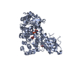 4b2gS S: Starting model for refinement |
|---|---|
| Similar structure data |


-Links
 PDBj
PDBj


- Assembly
| Deposited unit | 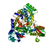
| ||||||||
|---|---|---|---|---|---|---|---|---|---|
| 1 |
| ||||||||
| Unit cell |
|
-Components
| #1: Protein | Mass: 66252.508 Da / Num. of mol.: 1 Source method: isolated from a genetically manipulated source Source: (gene. exp.) Oryza sativa subsp. indica (long-grained rice)Gene: GH3.8, OsI_025789 Production host:  Escherichia coli 'BL21-Gold(DE3)pLysS AG' (bacteria) Escherichia coli 'BL21-Gold(DE3)pLysS AG' (bacteria)References: UniProt: A3BLS0, Ligases; Forming carbon-nitrogen bonds; Acid-amino-acid ligases (peptide synthases) |
|---|---|
| #2: Chemical | ChemComp-AMP / Adenosine monophosphate  Mass: 347.221 Da / Num. of mol.: 1 / Source method: obtained synthetically / Formula: C10H14N5O7P / Feature type: SUBJECT OF INVESTIGATION / Comment: AMP*YM Mass: 347.221 Da / Num. of mol.: 1 / Source method: obtained synthetically / Formula: C10H14N5O7P / Feature type: SUBJECT OF INVESTIGATION / Comment: AMP*YM |
| #3: Water | ChemComp-HOH / Water Mass: 18.015 Da / Num. of mol.: 183 / Source method: isolated from a natural source / Formula: H2O Mass: 18.015 Da / Num. of mol.: 183 / Source method: isolated from a natural source / Formula: H2O |
| Has ligand of interest | Y |
-Experimental details
-Experiment
| Experiment | Method: X-RAY DIFFRACTION / Number of used crystals: 1 |
|---|
- Sample preparation
| Crystal | Density Matthews: 2 Å3/Da / Density % sol: 38.52 % |
|---|---|
| Crystal grow | Temperature: 289 K / Method: vapor diffusion, hanging drop Details: 0.08M Tris pH 8.0, 0.1M Tris-HCl (pH 8.5), 22.4% (w/v) PEG 4000, 0.02M Bis-Tris pH 6.5, 5% (w/v) PEG 3350, 0.2 Microliter 275.0mM 2,6-Dimethyl-4-heptyl-beta-D-maltopyranoside |
-Data collection
| Diffraction | Mean temperature: 100 K / Serial crystal experiment: N |
|---|---|
| Diffraction source | Source: SYNCHROTRON / Site: SSRF / Beamline: BL17U1 / Wavelength: 0.979183 Å |
| Detector | Type: PSI PILATUS 6M / Detector: PIXEL / Date: Jan 13, 2020 |
| Radiation | Protocol: MAD / Monochromatic (M) / Laue (L): M / Scattering type: x-ray |
| Radiation wavelength | Wavelength: 0.979183 Å / Relative weight: 1 |
| Reflection | Resolution: 1.99→30.5203 Å / Num. obs: 35491 / % possible obs: 98.44 % / Redundancy: 19.8 % / Rmerge(I) obs: 0.02218 / Net I/σ(I): 20.3 |
| Reflection shell | Resolution: 1.99→2.04 Å / Rmerge(I) obs: 0.3072 / Num. unique obs: 36047 |
- Processing
| Software |
| ||||||||||||||||||||||||||||||||||||||||||||||||||||||||||||||||||||||||||||||||||||||||||
|---|---|---|---|---|---|---|---|---|---|---|---|---|---|---|---|---|---|---|---|---|---|---|---|---|---|---|---|---|---|---|---|---|---|---|---|---|---|---|---|---|---|---|---|---|---|---|---|---|---|---|---|---|---|---|---|---|---|---|---|---|---|---|---|---|---|---|---|---|---|---|---|---|---|---|---|---|---|---|---|---|---|---|---|---|---|---|---|---|---|---|---|
| Refinement | Method to determine structure: MOLECULAR REPLACEMENT Starting model: 4b2g Resolution: 1.99→24.578 Å / SU ML: 0.26 / Cross valid method: THROUGHOUT / σ(F): 0 / Phase error: 26.76 / Stereochemistry target values: ML
| ||||||||||||||||||||||||||||||||||||||||||||||||||||||||||||||||||||||||||||||||||||||||||
| Solvent computation | Shrinkage radii: 0.9 Å / VDW probe radii: 1.11 Å / Solvent model: FLAT BULK SOLVENT MODEL | ||||||||||||||||||||||||||||||||||||||||||||||||||||||||||||||||||||||||||||||||||||||||||
| Displacement parameters | Biso max: 89.62 Å2 / Biso mean: 47.5662 Å2 / Biso min: 26 Å2 | ||||||||||||||||||||||||||||||||||||||||||||||||||||||||||||||||||||||||||||||||||||||||||
| Refinement step | Cycle: final / Resolution: 1.99→24.578 Å
| ||||||||||||||||||||||||||||||||||||||||||||||||||||||||||||||||||||||||||||||||||||||||||
| LS refinement shell | Refine-ID: X-RAY DIFFRACTION / Rfactor Rfree error: 0
|
