 Movie
Movie Controller

[English] 日本語
 Yorodumi
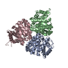
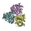
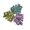
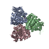
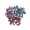
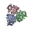
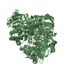
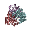
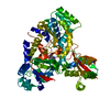
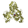
- PDB-3hpd: Structure of hydroxyethylthiazole kinase protein from pyrococcus ... -
+ Open data

- Basic information
| Entry | Database: PDB / ID: 3hpd | |||||||||
|---|---|---|---|---|---|---|---|---|---|---|
| Title | Structure of hydroxyethylthiazole kinase protein from pyrococcus horikoshii OT3 | |||||||||
 Components Components | Hydroxyethylthiazole kinase | |||||||||
 Keywords Keywords | TRANSFERASE / ALPHA-BETA / ATP BINDING / KINASE / ATP-binding / Magnesium / Metal-binding / Nucleotide-binding / Thiamine biosynthesis / Structural Genomics / NPPSFA / National Project on Protein Structural and Functional Analyses / RIKEN Structural Genomics/Proteomics Initiative / RSGI | |||||||||
| Function / homology |  Function and homology informationhydroxyethylthiazole kinase / hydroxyethylthiazole kinase activity / thiamine diphosphate biosynthetic process / thiamine biosynthetic process / magnesium ion binding / ATP binding Function and homology informationhydroxyethylthiazole kinase / hydroxyethylthiazole kinase activity / thiamine diphosphate biosynthetic process / thiamine biosynthetic process / magnesium ion binding / ATP bindingSimilarity search - Function | |||||||||
| Biological species |   Pyrococcus horikoshii (archaea) Pyrococcus horikoshii (archaea) | |||||||||
| Method | X-RAY DIFFRACTION / SYNCHROTRON / MOLECULAR REPLACEMENT / Resolution: 1.85 Å | |||||||||
 Authors Authors | Jeyakanthan, J. / Thamotharan, S. / Kuramitsu, S. / Yokoyama, S. / Tahirov, T.H. / RIKEN Structural Genomics/Proteomics Initiative (RSGI) | |||||||||
 Citation Citation | Journal: To be Published Title: Structure of Hydroxyethylthiazole Kinase Protein from Pyrococcus Horikoshii Ot3 Authors: Jeyakanthan, J. / Kuramitsu, S. / Yokoyama, S. | |||||||||
| History |
|
- Structure visualization
| Structure viewer | Molecule: MolmilJmol/JSmol |
|---|
- Downloads & links
-Download
| PDBx/mmCIF format | 3hpd.cif.gz | 66.2 KB | Display | PDBx/mmCIF format |
|---|---|---|---|---|
| PDB format | pdb3hpd.ent.gz | 48.5 KB | Display | PDB format |
| PDBx/mmJSON format | 3hpd.json.gz | Tree view | PDBx/mmJSON format | |
| Others |  Other downloads Other downloads |
-Validation report
| Arichive directory | https://data.pdbj.org/pub/pdb/validation_reports/hp/3hpdftp://data.pdbj.org/pub/pdb/validation_reports/hp/3hpd | HTTPS FTP |
|---|
-Related structure data
| Related structure data | 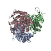 1c3qS S: Starting model for refinement |
|---|---|
| Similar structure data | |
| Other databases |
-Links
 PDBj
PDBj- Assembly
| Deposited unit | 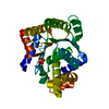
| ||||||||||||||||||
|---|---|---|---|---|---|---|---|---|---|---|---|---|---|---|---|---|---|---|---|
| 1 | 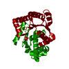
| ||||||||||||||||||
| Unit cell |
| ||||||||||||||||||
| Components on special symmetry positions |
|
-Components
| #1: Protein | / 4-methyl-5-beta-hydroxyethylthiazole kinase / Thz kinase / TH kinase Mass: 28990.275 Da / Num. of mol.: 1 Source method: isolated from a genetically manipulated source Source: (gene. exp.) Pyrococcus horikoshii (archaea) / Strain: OT3 / Plasmid: PET-11A / Production host:  Escherichia coli (E. coli) / Strain (production host): BL21-CODON PLUS(DE3)-RIL / References: UniProt: O58877, hydroxyethylthiazole kinase Escherichia coli (E. coli) / Strain (production host): BL21-CODON PLUS(DE3)-RIL / References: UniProt: O58877, hydroxyethylthiazole kinase |
|---|---|
| #2: Chemical | ChemComp-PO4 / Phosphate  Mass: 94.971 Da / Num. of mol.: 1 / Source method: obtained synthetically / Formula: PO4 Mass: 94.971 Da / Num. of mol.: 1 / Source method: obtained synthetically / Formula: PO4 |
| #3: Water | ChemComp-HOH / Water Mass: 18.015 Da / Num. of mol.: 208 / Source method: isolated from a natural source / Formula: H2O Mass: 18.015 Da / Num. of mol.: 208 / Source method: isolated from a natural source / Formula: H2O |
-Experimental details
-Experiment
| Experiment | Method: X-RAY DIFFRACTION / Number of used crystals: 1 |
|---|
- Sample preparation
| Crystal | Density Matthews: 2.03 Å3/Da / Density % sol: 39.48 % |
|---|---|
| Crystal grow | Temperature: 295 K / Method: microbatch method / pH: 9.3 Details: 0.88M SODIUMCITRATE, 0.1M CHESS, pH 9.3, MICROBATCH METHOD, temperature 295K |
-Data collection
| Diffraction | Mean temperature: 100 K |
|---|---|
| Diffraction source | Source: SYNCHROTRON / Site: SPring-8  / Beamline: BL26B1 / Wavelength: 1 Å / Beamline: BL26B1 / Wavelength: 1 Å |
| Detector | Type: RIGAKU RAXIS / Detector: IMAGE PLATE / Date: Nov 27, 2003 / Details: Mirrors |
| Radiation | Monochromator: Si (1 1 1), GRAPHITE / Protocol: SINGLE WAVELENGTH / Monochromatic (M) / Laue (L): M / Scattering type: x-ray |
| Radiation wavelength | Wavelength: 1 Å / Relative weight: 1 |
| Reflection | Resolution: 1.85→19.84 Å / Num. obs: 20162 / % possible obs: 99.8 % / Biso Wilson estimate: 11.3 Å2 / Rmerge(I) obs: 0.056 / Rsym value: 0.067 |
| Reflection shell | Resolution: 1.85→1.92 Å / Rmerge(I) obs: 0.324 / Num. unique all: 2004 / Rsym value: 0.292 / % possible all: 99.5 |
- Processing
| Software |
| ||||||||||||||||||||||||||||||||||||
|---|---|---|---|---|---|---|---|---|---|---|---|---|---|---|---|---|---|---|---|---|---|---|---|---|---|---|---|---|---|---|---|---|---|---|---|---|---|
| Refinement | Method to determine structure: MOLECULAR REPLACEMENT Starting model: 1C3Q Resolution: 1.85→19.84 Å / Rfactor Rfree error: 0.006 / Data cutoff high absF: 853468.49 / Data cutoff low absF: 0 / Isotropic thermal model: RESTRAINED / Cross valid method: THROUGHOUT / σ(F): 0 / Stereochemistry target values: Engh & Huber
| ||||||||||||||||||||||||||||||||||||
| Solvent computation | Solvent model: FLAT MODEL / Bsol: 57.8931 Å2 / ksol: 0.402666 e/Å3 | ||||||||||||||||||||||||||||||||||||
| Displacement parameters | Biso mean: 19.1 Å2 | ||||||||||||||||||||||||||||||||||||
| Refine analyze |
| ||||||||||||||||||||||||||||||||||||
| Refinement step | Cycle: LAST / Resolution: 1.85→19.84 Å
| ||||||||||||||||||||||||||||||||||||
| Refine LS restraints |
| ||||||||||||||||||||||||||||||||||||
| LS refinement shell | Resolution: 1.85→1.97 Å / Rfactor Rfree error: 0.017 / Total num. of bins used: 6
| ||||||||||||||||||||||||||||||||||||
| Xplor file |
|
