- PDB-7dig: Green fluorescent protein from Dendronephthya sp. SSAL-2002 -
+
Open data
ID or keywords:
Loading...
-
Basic information
Entry
Database: PDB / ID: 7dig
Title
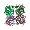
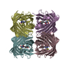
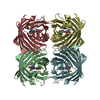
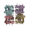
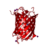
Green fluorescent protein from Dendronephthya sp. SSAL-2002
Components
Green fluorescent protein
Keywords
FLUORESCENT PROTEIN / Dendra / Dendronephthya / GFP
Function / homology
Green fluorescent protein, GFP / Green fluorescent protein-related / Green fluorescent protein / Green fluorescent protein / bioluminescence / generation of precursor metabolites and energy / metal ion binding / Green fluorescent protein
A: Green fluorescent protein B: Green fluorescent protein C: Green fluorescent protein D: Green fluorescent protein E: Green fluorescent protein F: Green fluorescent protein G: Green fluorescent protein H: Green fluorescent protein
Resolution: 2.6→48.7 Å / Cor.coef. Fo:Fc: 0.92 / Cor.coef. Fo:Fc free: 0.882 / SU B: 17.242 / SU ML: 0.347 / Cross valid method: THROUGHOUT / σ(F): 0 / ESU R Free: 0.394 / Stereochemistry target values: MAXIMUM LIKELIHOOD Details: HYDROGENS HAVE BEEN ADDED IN THE RIDING POSITIONS U VALUES : REFINED INDIVIDUALLY
Rfactor
Num. reflection
% reflection
Selection details
Rfree
0.2813
2707
4.9 %
RANDOM
Rwork
0.2342
-
-
-
obs
0.2364
52498
95.07 %
-
Solvent computation
Ion probe radii: 0.8 Å / Shrinkage radii: 0.8 Å / VDW probe radii: 1.2 Å / Solvent model: MASK
In the structure databanks used in Yorodumi, some data are registered as the other names, "COVID-19 virus" and "2019-nCoV". Here are the details of the virus and the list of structure data.
Jan 31, 2019. EMDB accession codes are about to change! (news from PDBe EMDB page)
EMDB accession codes are about to change! (news from PDBe EMDB page)
The allocation of 4 digits for EMDB accession codes will soon come to an end. Whilst these codes will remain in use, new EMDB accession codes will include an additional digit and will expand incrementally as the available range of codes is exhausted. The current 4-digit format prefixed with “EMD-” (i.e. EMD-XXXX) will advance to a 5-digit format (i.e. EMD-XXXXX), and so on. It is currently estimated that the 4-digit codes will be depleted around Spring 2019, at which point the 5-digit format will come into force.
The EM Navigator/Yorodumi systems omit the EMD- prefix.
Related info.:Q: What is EMD? / ID/Accession-code notation in Yorodumi/EM Navigator
Yorodumi is a browser for structure data from EMDB, PDB, SASBDB, etc.
This page is also the successor to EM Navigator detail page, and also detail information page/front-end page for Omokage search.
The word "yorodu" (or yorozu) is an old Japanese word meaning "ten thousand". "mi" (miru) is to see.
Related info.:EMDB / PDB / SASBDB / Comparison of 3 databanks / Yorodumi Search / Aug 31, 2016. New EM Navigator & Yorodumi / Yorodumi Papers / Jmol/JSmol / Function and homology information / Changes in new EM Navigator and Yorodumi
 Movie
Movie Controller


 Open data
 Basic information
 Components
 Keywords
 Function and homology information
 Dendronephthya sp. SSAL-2002 (invertebrata)
 Authors
 Korea, Republic Of, 1items
  Citation
 Structure visualization
 Downloads & links
 Other downloads
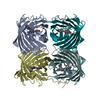


 PDBj
PDBj

 Assembly


 Sample preparation
 Processing