 Movie
Movie Controller

[English] 日本語
 Yorodumi
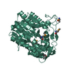
- PDB-7dg5: Crystal structure of mouse Smc1-Smc3 hinge domain containing a D5... -
+ Open data

- Basic information
| Entry | Database: PDB / ID: 7dg5 | ||||||
|---|---|---|---|---|---|---|---|
| Title | Crystal structure of mouse Smc1-Smc3 hinge domain containing a D574Y mutation | ||||||
 Components Components |
| ||||||
 Keywords Keywords |  DNA BINDING PROTEIN / cohesin / Smc1 / Smc3 / hinge DNA BINDING PROTEIN / cohesin / Smc1 / Smc3 / hinge | ||||||
| Function / homology |  Function and homology information Function and homology informationCohesin Loading onto Chromatin / Establishment of Sister Chromatid Cohesion / response to DNA damage checkpoint signaling / SUMOylation of DNA damage response and repair proteins / cohesin loader activity / meiotic cohesin complex / mitotic cohesin complex / cohesin complex / Resolution of Sister Chromatid Cohesion / Separation of Sister Chromatids ...Cohesin Loading onto Chromatin / Establishment of Sister Chromatid Cohesion / response to DNA damage checkpoint signaling / SUMOylation of DNA damage response and repair proteins / cohesin loader activity / meiotic cohesin complex / mitotic cohesin complex / cohesin complex / Resolution of Sister Chromatid Cohesion / Separation of Sister Chromatids / synaptonemal complex / lateral element / mediator complex binding / sister chromatid cohesion / mitotic sister chromatid cohesion / stem cell population maintenance / dynein complex binding / beta-tubulin binding / mitotic spindle pole / regulation of DNA replication / somatic stem cell population maintenance / chromosome, centromeric region / basement membrane / mitotic spindle assembly / meiotic cell cycle / response to radiation / kinetochore / nuclear matrix / double-stranded DNA binding / protein heterodimerization activity / cell division / DNA repair / chromatin binding / chromatin / ATP hydrolysis activity / DNA binding / nucleoplasm / ATP binding / nucleus / cytosolSimilarity search - Function | ||||||
| Biological species |  Mus musculus (house mouse) Mus musculus (house mouse) | ||||||
| Method | X-RAY DIFFRACTION / SYNCHROTRON / MOLECULAR REPLACEMENT / Resolution: 2 Å | ||||||
 Authors Authors | Seo, H. / Noh, H. / Oh, B.-H. | ||||||
| Funding support |  Korea, Republic Of, 1items Korea, Republic Of, 1items
| ||||||
 Citation Citation | Journal: Elife / Year: 2021 Title: Folding of cohesin's coiled coil is important for Scc2/4-induced association with chromosomes. Authors: Naomi J Petela / Andres Gonzalez Llamazares / Sarah Dixon / Bin Hu / Byung-Gil Lee / Jean Metson / Heekyo Seo / Antonio Ferrer-Harding / Menelaos Voulgaris / Thomas Gligoris / James Collier ...Authors: Naomi J Petela / Andres Gonzalez Llamazares / Sarah Dixon / Bin Hu / Byung-Gil Lee / Jean Metson / Heekyo Seo / Antonio Ferrer-Harding / Menelaos Voulgaris / Thomas Gligoris / James Collier / Byung-Ha Oh / Jan Löwe / Kim A Nasmyth /  Abstract: Cohesin's association with and translocation along chromosomal DNAs depend on an ATP hydrolysis cycle driving the association and subsequent release of DNA. This involves DNA being 'clamped' by Scc2 ...Cohesin's association with and translocation along chromosomal DNAs depend on an ATP hydrolysis cycle driving the association and subsequent release of DNA. This involves DNA being 'clamped' by Scc2 and ATP-dependent engagement of cohesin's Smc1 and Smc3 head domains. Scc2's replacement by Pds5 abrogates cohesin's ATPase and has an important role in halting DNA loop extrusion. The ATPase domains of all SMC proteins are separated from their hinge dimerisation domains by 50-nm-long coiled coils, which have been observed to zip up along their entire length and fold around an elbow, thereby greatly shortening the distance between hinges and ATPase heads. Whether folding exists in vivo or has any physiological importance is not known. We present here a cryo-EM structure of the form of cohesin that reveals the structure of folded and zipped-up coils in unprecedented detail and shows that Scc2 can associate with Smc1's ATPase head even when it is fully disengaged from that of Smc3. Using cysteine-specific crosslinking, we show that cohesin's coiled coils are frequently folded in vivo, including when cohesin holds sister chromatids together. Moreover, we describe a mutation () within Smc1's hinge that alters how Scc2 and Pds5 interact with Smc1's hinge and that enables Scc2 to support loading in the absence of its normal partner Scc4. The mutant phenotype of loading without Scc4 is only explicable if loading depends on an association between Scc2/4 and cohesin's hinge, which in turn requires coiled coil folding. | ||||||
| History |
|
- Structure visualization
| Structure viewer | Molecule: MolmilJmol/JSmol |
|---|
- Downloads & links
-Download
| PDBx/mmCIF format | 7dg5.cif.gz | 166.3 KB | Display | PDBx/mmCIF format |
|---|---|---|---|---|
| PDB format | pdb7dg5.ent.gz | 126.5 KB | Display | PDB format |
| PDBx/mmJSON format | 7dg5.json.gz | Tree view | PDBx/mmJSON format | |
| Others |  Other downloads Other downloads |
-Validation report
| Arichive directory | https://data.pdbj.org/pub/pdb/validation_reports/dg/7dg5ftp://data.pdbj.org/pub/pdb/validation_reports/dg/7dg5 | HTTPS FTP |
|---|
-Related structure data
| Related structure data |  7ogtC 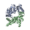 2wd5S S: Starting model for refinement C: citing same article ( |
|---|---|
| Similar structure data |


-Links
 PDBj
PDBj

- Assembly
| Deposited unit | 
| ||||||||
|---|---|---|---|---|---|---|---|---|---|
| 1 | 
| ||||||||
| 2 | 
| ||||||||
| Unit cell |
|
-Components
| #1: Protein | Mass: 24631.336 Da / Num. of mol.: 2 / Mutation: D574Y Source method: isolated from a genetically manipulated source Source: (gene. exp.) Mus musculus (house mouse) / Gene: Smc1a, Sb1.8, Smc1, Smc1l1, Smcb / Production host:  Escherichia coli K-12 (bacteria) / References: UniProt: Q9CU62 Escherichia coli K-12 (bacteria) / References: UniProt: Q9CU62#2: Protein | Mass: 24339.887 Da / Num. of mol.: 2 Source method: isolated from a genetically manipulated source Source: (gene. exp.) Mus musculus (house mouse) / Gene: Smc3, Bam, Bmh, Cspg6, Mmip1, Smc3l1, Smcd / Production host: Escherichia coli K-12 (bacteria) / References: UniProt: Q9CW03#3: Water | ChemComp-HOH / | Water Mass: 18.015 Da / Num. of mol.: 354 / Source method: isolated from a natural source / Formula: H2O Mass: 18.015 Da / Num. of mol.: 354 / Source method: isolated from a natural source / Formula: H2O |
|---|
-Experimental details
-Experiment
| Experiment | Method: X-RAY DIFFRACTION / Number of used crystals: 1 |
|---|
- Sample preparation
| Crystal | Density Matthews: 2.31 Å3/Da / Density % sol: 46.77 % |
|---|---|
| Crystal grow | Temperature: 295 K / Method: vapor diffusion, hanging drop / pH: 7 / Details: Sodium phosphate, Potassium phosphate, PEG 3350 |
-Data collection
| Diffraction | Mean temperature: 100 K / Serial crystal experiment: N | |||||||||||||||||||||||||||||||||||||||||||||||||||||||||||||||||||||||||||||||||||||||||||||||||||||||||||||||||||||||||||||||||||||||||||||||||||||||||||||||||||||||||||||||||||||||||||||
|---|---|---|---|---|---|---|---|---|---|---|---|---|---|---|---|---|---|---|---|---|---|---|---|---|---|---|---|---|---|---|---|---|---|---|---|---|---|---|---|---|---|---|---|---|---|---|---|---|---|---|---|---|---|---|---|---|---|---|---|---|---|---|---|---|---|---|---|---|---|---|---|---|---|---|---|---|---|---|---|---|---|---|---|---|---|---|---|---|---|---|---|---|---|---|---|---|---|---|---|---|---|---|---|---|---|---|---|---|---|---|---|---|---|---|---|---|---|---|---|---|---|---|---|---|---|---|---|---|---|---|---|---|---|---|---|---|---|---|---|---|---|---|---|---|---|---|---|---|---|---|---|---|---|---|---|---|---|---|---|---|---|---|---|---|---|---|---|---|---|---|---|---|---|---|---|---|---|---|---|---|---|---|---|---|---|---|---|---|---|---|
| Diffraction source | Source: SYNCHROTRON / Site: PAL/PLS / Beamline: 5C (4A) / Wavelength: 0.9796 Å | |||||||||||||||||||||||||||||||||||||||||||||||||||||||||||||||||||||||||||||||||||||||||||||||||||||||||||||||||||||||||||||||||||||||||||||||||||||||||||||||||||||||||||||||||||||||||||||
| Detector | Type: DECTRIS PILATUS3 6M / Detector: PIXEL / Date: May 12, 2016 | |||||||||||||||||||||||||||||||||||||||||||||||||||||||||||||||||||||||||||||||||||||||||||||||||||||||||||||||||||||||||||||||||||||||||||||||||||||||||||||||||||||||||||||||||||||||||||||
| Radiation | Protocol: SINGLE WAVELENGTH / Monochromatic (M) / Laue (L): M / Scattering type: x-ray | |||||||||||||||||||||||||||||||||||||||||||||||||||||||||||||||||||||||||||||||||||||||||||||||||||||||||||||||||||||||||||||||||||||||||||||||||||||||||||||||||||||||||||||||||||||||||||||
| Radiation wavelength | Wavelength: 0.9796 Å / Relative weight: 1 | |||||||||||||||||||||||||||||||||||||||||||||||||||||||||||||||||||||||||||||||||||||||||||||||||||||||||||||||||||||||||||||||||||||||||||||||||||||||||||||||||||||||||||||||||||||||||||||
| Reflection | Resolution: 2→50 Å / Num. obs: 52576 / % possible obs: 88.6 % / Redundancy: 2.6 % / Rmerge(I) obs: 0.064 / Rpim(I) all: 0.044 / Rrim(I) all: 0.078 / Χ2: 0.759 / Net I/σ(I): 7.3 / Num. measured all: 134894 | |||||||||||||||||||||||||||||||||||||||||||||||||||||||||||||||||||||||||||||||||||||||||||||||||||||||||||||||||||||||||||||||||||||||||||||||||||||||||||||||||||||||||||||||||||||||||||||
| Reflection shell | Diffraction-ID: 1
|
- Processing
| Software |
| |||||||||||||||||||||||||||||||||||||||||||||||||||||||||||||||||||||||||||||||||||||||||||||||||||||||||
|---|---|---|---|---|---|---|---|---|---|---|---|---|---|---|---|---|---|---|---|---|---|---|---|---|---|---|---|---|---|---|---|---|---|---|---|---|---|---|---|---|---|---|---|---|---|---|---|---|---|---|---|---|---|---|---|---|---|---|---|---|---|---|---|---|---|---|---|---|---|---|---|---|---|---|---|---|---|---|---|---|---|---|---|---|---|---|---|---|---|---|---|---|---|---|---|---|---|---|---|---|---|---|---|---|---|---|
| Refinement | Method to determine structure: MOLECULAR REPLACEMENT Starting model: 2WD5 Resolution: 2→42.06 Å / SU ML: 0.26 / Cross valid method: THROUGHOUT / σ(F): 2.18 / Phase error: 27.3 / Stereochemistry target values: ML
| |||||||||||||||||||||||||||||||||||||||||||||||||||||||||||||||||||||||||||||||||||||||||||||||||||||||||
| Solvent computation | Shrinkage radii: 0.9 Å / VDW probe radii: 1.11 Å / Solvent model: FLAT BULK SOLVENT MODEL | |||||||||||||||||||||||||||||||||||||||||||||||||||||||||||||||||||||||||||||||||||||||||||||||||||||||||
| Displacement parameters | Biso max: 60.69 Å2 / Biso mean: 20.6405 Å2 / Biso min: 5.15 Å2 | |||||||||||||||||||||||||||||||||||||||||||||||||||||||||||||||||||||||||||||||||||||||||||||||||||||||||
| Refinement step | Cycle: final / Resolution: 2→42.06 Å
| |||||||||||||||||||||||||||||||||||||||||||||||||||||||||||||||||||||||||||||||||||||||||||||||||||||||||
| LS refinement shell | Refine-ID: X-RAY DIFFRACTION / Rfactor Rfree error: 0 / Total num. of bins used: 14
|
