 Movie
Movie Controller


+ Open data

- Basic information
| Entry | Database: PDB / ID: 7d9y | ||||||
|---|---|---|---|---|---|---|---|
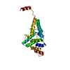
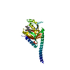
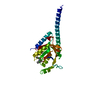
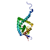
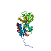
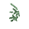
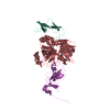
| Title | DNA binding domain of human DNA Ligase IV mutant - A3V | ||||||
 Components Components | DNA ligase 4 | ||||||
 Keywords Keywords | LIGASE / DBD mutant A3V / Native / ATP dependant ligase / hypomorphic mutant | ||||||
| Function / homology |  Function and homology information Function and homology informationDNA ligation involved in DNA recombination / positive regulation of chromosome organization / DNA ligase IV complex / DNA ligation involved in DNA repair / DNA ligase activity / DN2 thymocyte differentiation / DNA ligase (ATP) / T cell receptor V(D)J recombination / pro-B cell differentiation / DNA ligase (ATP) activity ...DNA ligation involved in DNA recombination / positive regulation of chromosome organization / DNA ligase IV complex / DNA ligation involved in DNA repair / DNA ligase activity / DN2 thymocyte differentiation / DNA ligase (ATP) / T cell receptor V(D)J recombination / pro-B cell differentiation / DNA ligase (ATP) activity / DNA-dependent protein kinase-DNA ligase 4 complex / single strand break repair / immunoglobulin V(D)J recombination / nonhomologous end joining complex / DNA ligation / V(D)J recombination / double-strand break repair via classical nonhomologous end joining / isotype switching / nucleotide-excision repair, DNA gap filling / positive regulation of neurogenesis / DNA biosynthetic process / cellular response to lithium ion / 2-LTR circle formation / somatic stem cell population maintenance / ligase activity / response to X-ray / chromosome organization / condensed chromosome / neurogenesis / central nervous system development / stem cell proliferation / cellular response to ionizing radiation / response to gamma radiation / Nonhomologous End-Joining (NHEJ) / double-strand break repair via nonhomologous end joining / establishment of integrated proviral latency / double-strand break repair / positive regulation of fibroblast proliferation / T cell differentiation in thymus / fibroblast proliferation / neuron apoptotic process / in utero embryonic development / negative regulation of neuron apoptotic process / cell population proliferation / chromosome, telomeric region / cell cycle / cell division / intracellular membrane-bounded organelle / magnesium ion binding / DNA binding / nucleoplasm / ATP binding / nucleusSimilarity search - Function | ||||||
| Biological species |  Homo sapiens (human) Homo sapiens (human) | ||||||
| Method | X-RAY DIFFRACTION / SYNCHROTRON / MOLECULAR REPLACEMENT / Resolution: 2.76 Å | ||||||
 Authors Authors | Maddi, E.R. / Raghavan, S.C. / Natesh, R. | ||||||
| Funding support |  India, 1items India, 1items
| ||||||
 Citation Citation | Journal: Protein Eng.Des.Sel. / Year: 2021 Title: Hypomorphic mutations in human DNA ligase IV lead to compromised DNA binding efficiency, hydrophobicity and thermal stability. Authors: Maddi, E.R. / Raghavan, S.C. / Natesh, R. | ||||||
| History |
|
- Structure visualization
| Structure viewer | Molecule: MolmilJmol/JSmol |
|---|
- Downloads & links
-Download
| PDBx/mmCIF format | 7d9y.cif.gz | 58.5 KB | Display | PDBx/mmCIF format |
|---|---|---|---|---|
| PDB format | pdb7d9y.ent.gz | 42.5 KB | Display | PDB format |
| PDBx/mmJSON format | 7d9y.json.gz | Tree view | PDBx/mmJSON format | |
| Others |  Other downloads Other downloads |
-Validation report
| Arichive directory | https://data.pdbj.org/pub/pdb/validation_reports/d9/7d9yftp://data.pdbj.org/pub/pdb/validation_reports/d9/7d9y | HTTPS FTP |
|---|
-Related structure data
| Related structure data |  7d9kSC S: Starting model for refinement C: citing same article ( |
|---|---|
| Similar structure data |

-Links
 PDBj
PDBj


- Assembly
| Deposited unit | 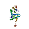
| ||||||||
|---|---|---|---|---|---|---|---|---|---|
| 1 |
| ||||||||
| Unit cell |
|
-Components
| #1: Protein | / DNA ligase IV / Polydeoxyribonucleotide synthase [ATP] 4 Mass: 27655.990 Da / Num. of mol.: 1 / Mutation: A3V Source method: isolated from a genetically manipulated source Source: (gene. exp.) Homo sapiens (human) / Gene: LIG4 / Variant: Natural variant VAR 029352 / Plasmid: pENH240_A3V, pET15bDetails (production host): Site Directed Mutagenesis at nucleotide position 8 on pENH240 Production host:  Escherichia coli (E. coli) / Strain (production host): BL21DE3 Codon Plus RIPL / References: UniProt: P49917, DNA ligase (ATP) Escherichia coli (E. coli) / Strain (production host): BL21DE3 Codon Plus RIPL / References: UniProt: P49917, DNA ligase (ATP) |
|---|---|
| #2: Water | ChemComp-HOH / Water Mass: 18.015 Da / Num. of mol.: 17 / Source method: isolated from a natural source / Formula: H2O Mass: 18.015 Da / Num. of mol.: 17 / Source method: isolated from a natural source / Formula: H2O |
-Experimental details
-Experiment
| Experiment | Method: X-RAY DIFFRACTION / Number of used crystals: 1 |
|---|
- Sample preparation
| Crystal | Density Matthews: 3.26 Å3/Da / Density % sol: 62.27 % Description: Cubic crystals of DBD LigIV mutant - A3V exhibiting a characteristic bipyramidal shape. Actual dimensions of the largest crystal are of the order of 0.25 mm to 0.3 mm. |
|---|---|
| Crystal grow | Temperature: 277.15 K / Method: vapor diffusion, hanging drop / pH: 8 Details: 0.125 M Tris pH 8.0, 20 % SOKALAN CP 42, 7.5% Methanol Temp details: Rubarth Vibration Free Incubator at 4 Deg Centigrade |
-Data collection
| Diffraction | Mean temperature: 100 K / Ambient temp details: LN2 / Serial crystal experiment: N | ||||||||||||||||||||||||
|---|---|---|---|---|---|---|---|---|---|---|---|---|---|---|---|---|---|---|---|---|---|---|---|---|---|
| Diffraction source | Source: SYNCHROTRON / Site: ESRF  / Beamline: ID30B / Wavelength: 0.97925 Å / Beamline: ID30B / Wavelength: 0.97925 Å | ||||||||||||||||||||||||
| Detector | Type: DECTRIS PILATUS3 S 6M / Detector: PIXEL / Date: Nov 22, 2018 / Details: Beamline Optics OH1/OH3 | ||||||||||||||||||||||||
| Radiation | Monochromator: Si (111) / Protocol: SINGLE WAVELENGTH / Monochromatic (M) / Laue (L): M / Scattering type: x-ray | ||||||||||||||||||||||||
| Radiation wavelength | Wavelength: 0.97925 Å / Relative weight: 1 | ||||||||||||||||||||||||
| Reflection | Resolution: 2.76→39.38 Å / Num. obs: 9928 / % possible obs: 100 % / Redundancy: 16.6 % / Biso Wilson estimate: 66.17 Å2 / CC1/2: 0.998 / Rmerge(I) obs: 0.138 / Rpim(I) all: 0.035 / Rrim(I) all: 0.143 / Net I/σ(I): 11.8 | ||||||||||||||||||||||||
| Reflection shell | Diffraction-ID: 1
|
- Processing
| Software |
| ||||||||||||||||||||||||
|---|---|---|---|---|---|---|---|---|---|---|---|---|---|---|---|---|---|---|---|---|---|---|---|---|---|
| Refinement | Method to determine structure: MOLECULAR REPLACEMENT Starting model: 7D9K Resolution: 2.76→39.38 Å / SU ML: 0.32 / Cross valid method: THROUGHOUT / σ(F): 1.34 / Phase error: 25.56 / Stereochemistry target values: ML
| ||||||||||||||||||||||||
| Solvent computation | Shrinkage radii: 0.9 Å / VDW probe radii: 1.11 Å / Solvent model: FLAT BULK SOLVENT MODEL | ||||||||||||||||||||||||
| Displacement parameters | Biso max: 150.3 Å2 / Biso mean: 66.7 Å2 / Biso min: 34.17 Å2 | ||||||||||||||||||||||||
| Refinement step | Cycle: final / Resolution: 2.76→39.38 Å
| ||||||||||||||||||||||||
| Refine LS restraints |
| ||||||||||||||||||||||||
| LS refinement shell | Refine-ID: X-RAY DIFFRACTION / Rfactor Rfree error: 0
|
