- PDB-7d9k: DNA binding domain of human DNA Ligase IV - Wild type -
+
Open data
ID or keywords:
Loading...
-
Basic information
Entry
Database: PDB / ID: 7d9k
Title
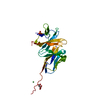
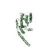
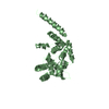
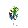
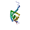
DNA binding domain of human DNA Ligase IV - Wild type
Components
DNA ligase 4
Keywords
LIGASE / DBD / Native / ATP dependant ligase.
Function / homology
Function and homology information
DNA ligation involved in DNA recombination / positive regulation of chromosome organization / DNA ligase IV complex / DNA ligation involved in DNA repair / DNA ligase activity / DN2 thymocyte differentiation / DNA ligase (ATP) / T cell receptor V(D)J recombination / pro-B cell differentiation / DNA ligase (ATP) activity ...DNA ligation involved in DNA recombination / positive regulation of chromosome organization / DNA ligase IV complex / DNA ligation involved in DNA repair / DNA ligase activity / DN2 thymocyte differentiation / DNA ligase (ATP) / T cell receptor V(D)J recombination / pro-B cell differentiation / DNA ligase (ATP) activity / DNA-dependent protein kinase-DNA ligase 4 complex / single strand break repair / immunoglobulin V(D)J recombination / nonhomologous end joining complex / DNA ligation / V(D)J recombination / double-strand break repair via classical nonhomologous end joining / isotype switching / nucleotide-excision repair, DNA gap filling / positive regulation of neurogenesis / DNA biosynthetic process / cellular response to lithium ion / 2-LTR circle formation / somatic stem cell population maintenance / ligase activity / response to X-ray / chromosome organization / condensed chromosome / neurogenesis / central nervous system development / stem cell proliferation / cellular response to ionizing radiation / response to gamma radiation / Nonhomologous End-Joining (NHEJ) / double-strand break repair via nonhomologous end joining / establishment of integrated proviral latency / double-strand break repair / positive regulation of fibroblast proliferation / T cell differentiation in thymus / fibroblast proliferation / neuron apoptotic process / in utero embryonic development / negative regulation of neuron apoptotic process / cell population proliferation / chromosome, telomeric region / cell cycle / cell division / intracellular membrane-bounded organelle / magnesium ion binding / DNA binding / nucleoplasm / ATP binding / nucleus Similarity search - Function
DNA ligase IV domain / DNA ligase IV / DNA ligase 4 / DNA Ligase 4, adenylation domain / DNA ligase, ATP-dependent / DNA ligase, ATP-dependent, N-terminal / DNA ligase, ATP-dependent, N-terminal domain superfamily / DNA ligase N terminus / ATP-dependent DNA ligase signature 2. / ATP-dependent DNA ligase AMP-binding site. ...DNA ligase IV domain / DNA ligase IV / DNA ligase 4 / DNA Ligase 4, adenylation domain / DNA ligase, ATP-dependent / DNA ligase, ATP-dependent, N-terminal / DNA ligase, ATP-dependent, N-terminal domain superfamily / DNA ligase N terminus / ATP-dependent DNA ligase signature 2. / ATP-dependent DNA ligase AMP-binding site. / DNA ligase, ATP-dependent, C-terminal / ATP dependent DNA ligase C terminal region / DNA ligase, ATP-dependent, conserved site / ATP-dependent DNA ligase family profile. / DNA ligase, ATP-dependent, central / ATP dependent DNA ligase domain / BRCA1 C Terminus (BRCT) domain / breast cancer carboxy-terminal domain / BRCT domain profile. / BRCT domain / BRCT domain superfamily / Nucleic acid-binding, OB-fold Similarity search - Domain/homology
DNAligase4 / / DNA ligase IV / Polydeoxyribonucleotide synthase [ATP] 4
Mass: 27627.938 Da / Num. of mol.: 1 / Fragment: DNA binding domain Source method: isolated from a genetically manipulated source Source: (gene. exp.) Homo sapiens (human) / Gene: LIG4 / Plasmid: pET15b, pENH240 Details (production host): DBDLigIV cloned at NdeI and BamHI restriction site in pET15b Production host: Escherichia coli (E. coli) / Strain (production host): BL21DE3 Codon Plus RIPL / References: UniProt: P49917, DNA ligase (ATP)
Mass: 18.015 Da / Num. of mol.: 21 / Source method: isolated from a natural source / Formula: H2O
-
Experimental details
-
Experiment
Experiment
Method: X-RAY DIFFRACTION / Number of used crystals: 1
-
Sample preparation
Crystal
Density Matthews: 3.18 Å3/Da / Density % sol: 61.33 % Description: Cubic crystals of DBD LigIV-Wt exhibiting a characteristic bipyramidal shape. Actual dimensions of the largest crystal are of the order of 0.25 um to 0.26 mm.
Crystal grow
Temperature: 277.15 K / Method: vapor diffusion, hanging drop / pH: 8 / Details: 0.1 M Tris pH 8.0, 20 % SOKALAN CP 42, 5% Methanol / Temp details: Rubarth Incubator Vibration Free
-
Data collection
Diffraction
Mean temperature: 100 K / Ambient temp details: LN2 / Serial crystal experiment: N
In the structure databanks used in Yorodumi, some data are registered as the other names, "COVID-19 virus" and "2019-nCoV". Here are the details of the virus and the list of structure data.
Jan 31, 2019. EMDB accession codes are about to change! (news from PDBe EMDB page)
EMDB accession codes are about to change! (news from PDBe EMDB page)
The allocation of 4 digits for EMDB accession codes will soon come to an end. Whilst these codes will remain in use, new EMDB accession codes will include an additional digit and will expand incrementally as the available range of codes is exhausted. The current 4-digit format prefixed with “EMD-” (i.e. EMD-XXXX) will advance to a 5-digit format (i.e. EMD-XXXXX), and so on. It is currently estimated that the 4-digit codes will be depleted around Spring 2019, at which point the 5-digit format will come into force.
The EM Navigator/Yorodumi systems omit the EMD- prefix.
Related info.:Q: What is EMD? / ID/Accession-code notation in Yorodumi/EM Navigator
Yorodumi is a browser for structure data from EMDB, PDB, SASBDB, etc.
This page is also the successor to EM Navigator detail page, and also detail information page/front-end page for Omokage search.
The word "yorodu" (or yorozu) is an old Japanese word meaning "ten thousand". "mi" (miru) is to see.
Related info.:EMDB / PDB / SASBDB / Comparison of 3 databanks / Yorodumi Search / Aug 31, 2016. New EM Navigator & Yorodumi / Yorodumi Papers / Jmol/JSmol / Function and homology information / Changes in new EM Navigator and Yorodumi
 Movie
Movie Controller


 Open data
 Basic information
 Components
 Keywords
 Function and homology information
 Authors
 India, 1items
  Citation
 Structure visualization
 Downloads & links
 Other downloads
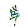


 PDBj
PDBj


 Assembly
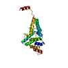

 Mass: 18.015 Da / Num. of mol.: 21 / Source method: isolated from a natural source / Formula: H2O
 Sample preparation
 / Beamline: ID30B / Wavelength: 0.97927 Å
 Processing