 Movie
Movie Controller


+ Open data

- Basic information
| Entry | Database: PDB / ID: 7d3d | ||||||
|---|---|---|---|---|---|---|---|
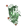
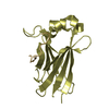
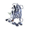
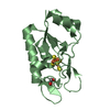
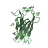
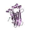
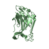
| Title | Crystal structure of SPOP bound with a peptide | ||||||
 Components Components |
| ||||||
 Keywords Keywords |  PROTEIN BINDING / ccRCC / E3 ligase adaptor SPOP / ubiquitination / SBC motif(F-pS-S/T-S/T / F is nonpolar / p is polar) PROTEIN BINDING / ccRCC / E3 ligase adaptor SPOP / ubiquitination / SBC motif(F-pS-S/T-S/T / F is nonpolar / p is polar) | ||||||
| Function / homology |  Function and homology informationregulation of proteolysis / Cul3-RING ubiquitin ligase complex / molecular function inhibitor activity / localization / Hedgehog 'on' state / protein polyubiquitination / proteasome-mediated ubiquitin-dependent protein catabolic process / nuclear speck / ubiquitin protein ligase binding / nucleoplasm ...regulation of proteolysis / Cul3-RING ubiquitin ligase complex / molecular function inhibitor activity / localization / Hedgehog 'on' state / protein polyubiquitination / proteasome-mediated ubiquitin-dependent protein catabolic process / nuclear speck / ubiquitin protein ligase binding / nucleoplasm / nucleus / cytoplasm Function and homology informationregulation of proteolysis / Cul3-RING ubiquitin ligase complex / molecular function inhibitor activity / localization / Hedgehog 'on' state / protein polyubiquitination / proteasome-mediated ubiquitin-dependent protein catabolic process / nuclear speck / ubiquitin protein ligase binding / nucleoplasm ...regulation of proteolysis / Cul3-RING ubiquitin ligase complex / molecular function inhibitor activity / localization / Hedgehog 'on' state / protein polyubiquitination / proteasome-mediated ubiquitin-dependent protein catabolic process / nuclear speck / ubiquitin protein ligase binding / nucleoplasm / nucleus / cytoplasmSimilarity search - Function | ||||||
| Biological species |  Homo sapiens (human) Homo sapiens (human) | ||||||
| Method | X-RAY DIFFRACTION / SYNCHROTRON / MOLECULAR REPLACEMENT / Resolution: 1.45 Å | ||||||
 Authors Authors | Yang, C.-G. / Gan, J.H. | ||||||
 Citation Citation | Journal: Chin.J.Chem. / Year: 2021 Title: A peptide binder of E3 ligase adaptor SPOP disrupts oncogenic SPOP-protein interactions in kidney cancer cells. Authors: Wang, Z. / Zhang, H. / Chen, B. / Ouyang, T. / Zheng, T. / Zhou, R. / Dong, Z. / Huang, Y. / Zhang, T. / Jiang, H. / Gan, J. / Luo, C. / Yang, C.-G. | ||||||
| History |
|
- Structure visualization
| Structure viewer | Molecule: MolmilJmol/JSmol |
|---|
- Downloads & links
-Download
| PDBx/mmCIF format | 7d3d.cif.gz | 148.8 KB | Display | PDBx/mmCIF format |
|---|---|---|---|---|
| PDB format | pdb7d3d.ent.gz | 116 KB | Display | PDB format |
| PDBx/mmJSON format | 7d3d.json.gz | Tree view | PDBx/mmJSON format | |
| Others |  Other downloads Other downloads |
-Validation report
| Arichive directory | https://data.pdbj.org/pub/pdb/validation_reports/d3/7d3dftp://data.pdbj.org/pub/pdb/validation_reports/d3/7d3d | HTTPS FTP |
|---|
-Related structure data
| Related structure data | 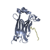 3ivvS S: Starting model for refinement |
|---|---|
| Similar structure data |


-Links
 PDBj
PDBj


- Assembly
| Deposited unit | 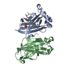
| ||||||||
|---|---|---|---|---|---|---|---|---|---|
| 1 | 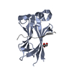
| ||||||||
| 2 | 
| ||||||||
| Unit cell |
|
-Components
| #1: Protein | Mass: 16141.605 Da / Num. of mol.: 2 / Fragment: UNP residues 28-166 Source method: isolated from a genetically manipulated source Source: (gene. exp.) Homo sapiens (human) / Gene: SPOP / Production host:  Escherichia coli (E. coli) / References: UniProt: O43791 Escherichia coli (E. coli) / References: UniProt: O43791#2: Protein/peptide | Mass: 1220.285 Da / Num. of mol.: 2 Source method: isolated from a genetically manipulated source Source: (gene. exp.) Homo sapiens (human) / Production host: Escherichia coli (E. coli)#3: Chemical | ChemComp-GOL / | Glycerol  Mass: 92.094 Da / Num. of mol.: 1 / Source method: obtained synthetically / Formula: C3H8O3 / Feature type: SUBJECT OF INVESTIGATION Mass: 92.094 Da / Num. of mol.: 1 / Source method: obtained synthetically / Formula: C3H8O3 / Feature type: SUBJECT OF INVESTIGATION#4: Water | ChemComp-HOH / | Water Mass: 18.015 Da / Num. of mol.: 363 / Source method: isolated from a natural source / Formula: H2O Mass: 18.015 Da / Num. of mol.: 363 / Source method: isolated from a natural source / Formula: H2OHas ligand of interest | Y | |
|---|
-Experimental details
-Experiment
| Experiment | Method: X-RAY DIFFRACTION / Number of used crystals: 1 |
|---|
- Sample preparation
| Crystal | Density Matthews: 2.03 Å3/Da / Density % sol: 39.49 % |
|---|---|
| Crystal grow | Temperature: 289 K / Method: vapor diffusion, sitting drop Details: 200 mM sodium acetate trihydrate, pH 7.0, 20%(w/v) polyethylene glycol 3350 |
-Data collection
| Diffraction | Mean temperature: 100 K / Serial crystal experiment: N | |||||||||||||||||||||||||||||||||||||||||||||||||||||||||||||||||||||||||||||||||||||||||||||||||||
|---|---|---|---|---|---|---|---|---|---|---|---|---|---|---|---|---|---|---|---|---|---|---|---|---|---|---|---|---|---|---|---|---|---|---|---|---|---|---|---|---|---|---|---|---|---|---|---|---|---|---|---|---|---|---|---|---|---|---|---|---|---|---|---|---|---|---|---|---|---|---|---|---|---|---|---|---|---|---|---|---|---|---|---|---|---|---|---|---|---|---|---|---|---|---|---|---|---|---|---|---|
| Diffraction source | Source: SYNCHROTRON / Site: SSRF  / Beamline: BL18U1 / Wavelength: 0.9735 Å / Beamline: BL18U1 / Wavelength: 0.9735 Å | |||||||||||||||||||||||||||||||||||||||||||||||||||||||||||||||||||||||||||||||||||||||||||||||||||
| Detector | Type: DECTRIS PILATUS 6M / Detector: PIXEL / Date: Feb 11, 2017 | |||||||||||||||||||||||||||||||||||||||||||||||||||||||||||||||||||||||||||||||||||||||||||||||||||
| Radiation | Protocol: SINGLE WAVELENGTH / Monochromatic (M) / Laue (L): M / Scattering type: x-ray | |||||||||||||||||||||||||||||||||||||||||||||||||||||||||||||||||||||||||||||||||||||||||||||||||||
| Radiation wavelength | Wavelength: 0.9735 Å / Relative weight: 1 | |||||||||||||||||||||||||||||||||||||||||||||||||||||||||||||||||||||||||||||||||||||||||||||||||||
| Reflection | Resolution: 1.44→49.73 Å / Num. obs: 50483 / % possible obs: 98.1 % / Redundancy: 11.9 % / Rmerge(I) obs: 0.073 / Rpim(I) all: 0.021 / Rrim(I) all: 0.076 / Χ2: 1.011 / Net I/σ(I): 8.4 | |||||||||||||||||||||||||||||||||||||||||||||||||||||||||||||||||||||||||||||||||||||||||||||||||||
| Reflection shell | Diffraction-ID: 1
|
- Processing
| Software |
| |||||||||||||||||||||||||||||||||||||||||||||||||||||||||||||||||||||||||||
|---|---|---|---|---|---|---|---|---|---|---|---|---|---|---|---|---|---|---|---|---|---|---|---|---|---|---|---|---|---|---|---|---|---|---|---|---|---|---|---|---|---|---|---|---|---|---|---|---|---|---|---|---|---|---|---|---|---|---|---|---|---|---|---|---|---|---|---|---|---|---|---|---|---|---|---|---|
| Refinement | Method to determine structure: MOLECULAR REPLACEMENT Starting model: 3IVV Resolution: 1.45→30 Å / Cor.coef. Fo:Fc: 0.966 / Cor.coef. Fo:Fc free: 0.957 / SU B: 2.153 / SU ML: 0.038 / Cross valid method: THROUGHOUT / σ(F): 0 / ESU R: 0.084 / ESU R Free: 0.068 / Stereochemistry target values: MAXIMUM LIKELIHOOD Details: HYDROGENS HAVE BEEN ADDED IN THE RIDING POSITIONS U VALUES : WITH TLS ADDED
| |||||||||||||||||||||||||||||||||||||||||||||||||||||||||||||||||||||||||||
| Solvent computation | Ion probe radii: 0.8 Å / Shrinkage radii: 0.8 Å / VDW probe radii: 1.2 Å / Solvent model: MASK | |||||||||||||||||||||||||||||||||||||||||||||||||||||||||||||||||||||||||||
| Displacement parameters | Biso max: 62.99 Å2 / Biso mean: 15.766 Å2 / Biso min: 6.21 Å2
| |||||||||||||||||||||||||||||||||||||||||||||||||||||||||||||||||||||||||||
| Refinement step | Cycle: final / Resolution: 1.45→30 Å
| |||||||||||||||||||||||||||||||||||||||||||||||||||||||||||||||||||||||||||
| Refine LS restraints |
| |||||||||||||||||||||||||||||||||||||||||||||||||||||||||||||||||||||||||||
| LS refinement shell | Resolution: 1.45→1.481 Å / Rfactor Rfree error: 0
| |||||||||||||||||||||||||||||||||||||||||||||||||||||||||||||||||||||||||||
| Refinement TLS params. | Method: refined / Refine-ID: X-RAY DIFFRACTION
| |||||||||||||||||||||||||||||||||||||||||||||||||||||||||||||||||||||||||||
| Refinement TLS group |
|
