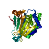
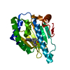
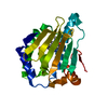
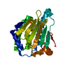
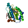
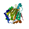
 Movie
Movie Controller


+ Open data

- Basic information
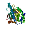
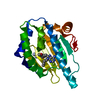
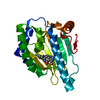
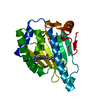
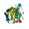
| Entry | Database: PDB / ID: 7d1v | ||||||
|---|---|---|---|---|---|---|---|
| Title | Hsp90 alpha N-terminal domain in complex with a 6C compund | ||||||
 Components Components | Heat shock protein HSP 90-alpha Heat shock response Heat shock response | ||||||
 Keywords Keywords | CHAPERONE/INHIBITOR / CHAPERONE-Inhibitor complex | ||||||
| Function / homology |  Function and homology information Function and homology informationDrug-mediated inhibition of ERBB2 signaling / ESR-mediated signaling / DDX58/IFIH1-mediated induction of interferon-alpha/beta / HSF1-dependent transactivation / Signaling by ERBB2 / Tetrahydrobiopterin (BH4) synthesis, recycling, salvage and regulation / eNOS activation / HSF1 activation / RHOBTB2 GTPase cycle / Sema3A PAK dependent Axon repulsion ...Drug-mediated inhibition of ERBB2 signaling / ESR-mediated signaling / DDX58/IFIH1-mediated induction of interferon-alpha/beta / HSF1-dependent transactivation / Signaling by ERBB2 / Tetrahydrobiopterin (BH4) synthesis, recycling, salvage and regulation / eNOS activation / HSF1 activation / RHOBTB2 GTPase cycle / Sema3A PAK dependent Axon repulsion / Downregulation of ERBB2 signaling / Attenuation phase / Regulation of necroptotic cell death / HSP90 chaperone cycle for steroid hormone receptors (SHR) in the presence of ligand / VEGFR2 mediated vascular permeability / positive regulation of cytotoxic T cell differentiation / Extra-nuclear estrogen signaling / The role of GTSE1 in G2/M progression after G2 checkpoint / Loss of Nlp from mitotic centrosomes / Recruitment of mitotic centrosome proteins and complexes / Loss of proteins required for interphase microtubule organization from the centrosome / Regulation of actin dynamics for phagocytic cup formation / Recruitment of NuMA to mitotic centrosomes / Anchoring of the basal body to the plasma membrane / AURKA Activation by TPX2 / Regulation of PLK1 Activity at G2/M Transition / Estrogen-dependent gene expression / VEGFA-VEGFR2 Pathway / sperm mitochondrial sheath / dATP binding / sulfonylurea receptor binding / CTP binding / positive regulation of protein polymerization / UTP binding / sperm plasma membrane / protein insertion into mitochondrial outer membrane / telomerase holoenzyme complex assembly / Rho GDP-dissociation inhibitor binding / TPR domain binding / non-chaperonin molecular chaperone ATPase / regulation of postsynaptic membrane neurotransmitter receptor levels / dendritic growth cone / skeletal muscle contraction / regulation of protein ubiquitination / positive regulation of cell size / telomere maintenance via telomerase / response to unfolded protein / chaperone-mediated protein complex assembly / sperm flagellum / positive regulation of lamellipodium assembly / DNA polymerase binding / axonal growth cone / protein folding chaperone / positive regulation of cardiac muscle contraction / cardiac muscle cell apoptotic process / response to salt stress / positive regulation of defense response to virus by host / protein tyrosine kinase binding / nitric oxide biosynthetic process / Neutrophil degranulation / response to cold / activation of innate immune response / positive regulation of interferon-beta production / nitric-oxide synthase regulator activity / response to cocaine / brush border membrane / ATP-dependent protein folding chaperone / neuron migration / tau protein binding / cellular response to virus / positive regulation of protein import into nucleus / response to estrogen / histone deacetylase binding / positive regulation of protein catabolic process / regulation of protein localization / positive regulation of nitric oxide biosynthetic process / disordered domain specific binding / unfolded protein binding / melanosome / protein folding / GTPase binding / myelin sheath / cellular response to heat / response to heat / protein refolding / scaffold protein binding / basolateral plasma membrane / protein phosphatase binding / collagen-containing extracellular matrix / regulation of apoptotic process / transmembrane transporter binding / protein stabilization / response to xenobiotic stimulus / positive regulation of protein phosphorylation / apical plasma membrane / response to antibiotic / mRNA binding / neuronal cell body / ubiquitin protein ligase binding / GTP bindingSimilarity search - Function | ||||||
| Biological species |  Mus musculus (house mouse) Mus musculus (house mouse) | ||||||
| Method | X-RAY DIFFRACTION / SYNCHROTRON / MOLECULAR REPLACEMENT / Resolution: 1.332 Å | ||||||
 Authors Authors | Shin, S.C. / Kim, E.E. | ||||||
| Funding support |  Korea, Republic Of, 1items Korea, Republic Of, 1items
| ||||||
 Citation Citation | Journal: Int J Mol Sci / Year: 2020 Title: Structural Basis for Design of New Purine-Based Inhibitors Targeting the Hydrophobic Binding Pocket of Hsp90. Authors: Shin, S.C. / El-Damasy, A.K. / Lee, J.H. / Seo, S.H. / Kim, J.H. / Seo, Y.H. / Lee, Y. / Yu, J.H. / Bang, E.K. / Kim, E.E. / Keum, G. | ||||||
| History |
|
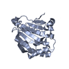
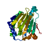
- Structure visualization
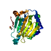
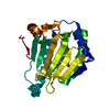
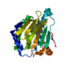
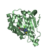
| Structure viewer | Molecule: MolmilJmol/JSmol |
|---|
- Downloads & links
-Download
| PDBx/mmCIF format | 7d1v.cif.gz | 68.1 KB | Display | PDBx/mmCIF format |
|---|---|---|---|---|
| PDB format | pdb7d1v.ent.gz | 46.6 KB | Display | PDB format |
| PDBx/mmJSON format | 7d1v.json.gz | Tree view | PDBx/mmJSON format | |
| Others |  Other downloads Other downloads |
-Validation report
| Arichive directory | https://data.pdbj.org/pub/pdb/validation_reports/d1/7d1vftp://data.pdbj.org/pub/pdb/validation_reports/d1/7d1v | HTTPS FTP |
|---|
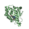
-Related structure data
| Related structure data |  7d22C  7d24C  7d25C 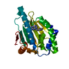 7d26C 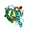 5h22S S: Starting model for refinement C: citing same article ( |
|---|---|
| Similar structure data |


-Links
 PDBj
PDBj


- Assembly
| Deposited unit | 
| ||||||||||
|---|---|---|---|---|---|---|---|---|---|---|---|
| 1 |
| ||||||||||
| Unit cell |
|
-Components
| #1: Protein | Heat shock response / Heat shock 86 kDa / HSP86 / Tumor-specific transplantation 86 kDa antigen / TSTA Mass: 24109.275 Da / Num. of mol.: 1 Source method: isolated from a genetically manipulated source Source: (gene. exp.) Mus musculus (house mouse) / Gene: Hsp90aa1, Hsp86, Hsp86-1, Hspca / Production host:  Escherichia coli (E. coli) / References: UniProt: P07901 Escherichia coli (E. coli) / References: UniProt: P07901 | ||||
|---|---|---|---|---|---|
| #2: Chemical | 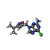  Mass: 292.724 Da / Num. of mol.: 2 / Source method: obtained synthetically / Formula: C12H13ClN6O / Feature type: SUBJECT OF INVESTIGATION Mass: 292.724 Da / Num. of mol.: 2 / Source method: obtained synthetically / Formula: C12H13ClN6O / Feature type: SUBJECT OF INVESTIGATION#3: Water | ChemComp-HOH / | Water Mass: 18.015 Da / Num. of mol.: 419 / Source method: isolated from a natural source / Formula: H2O Mass: 18.015 Da / Num. of mol.: 419 / Source method: isolated from a natural source / Formula: H2OHas ligand of interest | Y | |
-Experimental details
-Experiment
| Experiment | Method: X-RAY DIFFRACTION / Number of used crystals: 1 |
|---|
- Sample preparation
| Crystal | Density Matthews: 3.19 Å3/Da / Density % sol: 61.47 % |
|---|---|
| Crystal grow | Temperature: 295 K / Method: vapor diffusion, sitting drop / Details: 1.0M ammonium Sulfate, 0.1M Tris-HCl (pH8.5) |
-Data collection
| Diffraction | Mean temperature: 100 K / Serial crystal experiment: N | |||||||||||||||||||||||||||||||||||||||||||||||||||||||||||||||||||||||||||||
|---|---|---|---|---|---|---|---|---|---|---|---|---|---|---|---|---|---|---|---|---|---|---|---|---|---|---|---|---|---|---|---|---|---|---|---|---|---|---|---|---|---|---|---|---|---|---|---|---|---|---|---|---|---|---|---|---|---|---|---|---|---|---|---|---|---|---|---|---|---|---|---|---|---|---|---|---|---|---|
| Diffraction source | Source: SYNCHROTRON / Site: PAL/PLS / Beamline: 5C (4A) / Wavelength: 1 Å | |||||||||||||||||||||||||||||||||||||||||||||||||||||||||||||||||||||||||||||
| Detector | Type: ADSC QUANTUM 315r / Detector: CCD / Date: Feb 11, 2011 | |||||||||||||||||||||||||||||||||||||||||||||||||||||||||||||||||||||||||||||
| Radiation | Protocol: SINGLE WAVELENGTH / Monochromatic (M) / Laue (L): M / Scattering type: x-ray | |||||||||||||||||||||||||||||||||||||||||||||||||||||||||||||||||||||||||||||
| Radiation wavelength | Wavelength: 1 Å / Relative weight: 1 | |||||||||||||||||||||||||||||||||||||||||||||||||||||||||||||||||||||||||||||
| Reflection | Resolution: 1.33→50 Å / Num. obs: 68098 / % possible obs: 99.3 % / Redundancy: 10.8 % / Rmerge(I) obs: 0.056 / Χ2: 1.6 / Net I/σ(I): 15.9 / Num. measured all: 734331 | |||||||||||||||||||||||||||||||||||||||||||||||||||||||||||||||||||||||||||||
| Reflection shell |
|
- Processing
| Software |
| ||||||||||||||||||||||||||||||||||||||||||||||||||||||||||||||||||||||||||||||||||||
|---|---|---|---|---|---|---|---|---|---|---|---|---|---|---|---|---|---|---|---|---|---|---|---|---|---|---|---|---|---|---|---|---|---|---|---|---|---|---|---|---|---|---|---|---|---|---|---|---|---|---|---|---|---|---|---|---|---|---|---|---|---|---|---|---|---|---|---|---|---|---|---|---|---|---|---|---|---|---|---|---|---|---|---|---|---|
| Refinement | Method to determine structure: MOLECULAR REPLACEMENT Starting model: 5H22 Resolution: 1.332→29.959 Å / SU ML: 0.12 / Cross valid method: THROUGHOUT / σ(F): 1.5 / Phase error: 17.17 / Stereochemistry target values: ML
| ||||||||||||||||||||||||||||||||||||||||||||||||||||||||||||||||||||||||||||||||||||
| Solvent computation | Shrinkage radii: 0.9 Å / VDW probe radii: 1.11 Å / Solvent model: FLAT BULK SOLVENT MODEL | ||||||||||||||||||||||||||||||||||||||||||||||||||||||||||||||||||||||||||||||||||||
| Displacement parameters | Biso max: 50.03 Å2 / Biso mean: 17.972 Å2 / Biso min: 7.65 Å2 | ||||||||||||||||||||||||||||||||||||||||||||||||||||||||||||||||||||||||||||||||||||
| Refinement step | Cycle: final / Resolution: 1.332→29.959 Å
| ||||||||||||||||||||||||||||||||||||||||||||||||||||||||||||||||||||||||||||||||||||
| LS refinement shell | Refine-ID: X-RAY DIFFRACTION / Rfactor Rfree error: 0
|
