 Movie
Movie Controller

[English] 日本語
 Yorodumi
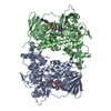
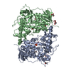
- PDB-7cu1: CRYSTAL STRUCTURE OF STREPTOMYCES ALBOGRISEOLUS FLAVIN-DEPENDENT ... -
+ Open data

- Basic information
| Entry | Database: PDB / ID: 7cu1 | ||||||
|---|---|---|---|---|---|---|---|
| Title | CRYSTAL STRUCTURE OF STREPTOMYCES ALBOGRISEOLUS FLAVIN-DEPENDENT TRYPTOPHAN 6-HALOGENASE (THAL) IN COMPLEX WITH FAD and AMP | ||||||
 Components Components | Tryptophan 6-halogenase | ||||||
 Keywords Keywords |  OXIDOREDUCTASE / FLAVIN MONOOXYGENASE / FLAVIN-DEPENDENT TRYPTOPHAN 6-HALOGENASE OXIDOREDUCTASE / FLAVIN MONOOXYGENASE / FLAVIN-DEPENDENT TRYPTOPHAN 6-HALOGENASE | ||||||
| Function / homology |  Function and homology information Function and homology information | ||||||
| Biological species |  Streptomyces albogriseolus (bacteria) Streptomyces albogriseolus (bacteria) | ||||||
| Method | X-RAY DIFFRACTION / MOLECULAR REPLACEMENT / Resolution: 1.91 Å | ||||||
| Model details | Crystal structure of flavin-dependent tryptophan 6-halogenase (Thal) in complex with FAD from ...Crystal structure of flavin-dependent tryptophan 6-halogenase (Thal) in complex with FAD from Streptomyces albogriseolus | ||||||
 Authors Authors | Chitnumsub, P. / Jaruwat, A. / Phintha, A. / Chaiyen, P. | ||||||
 Citation Citation | Journal: J.Biol.Chem. / Year: 2020 Title: Dissecting the low catalytic capability of flavin-dependent halogenases. Authors: Phintha, A. / Prakinee, K. / Jaruwat, A. / Lawan, N. / Visitsatthawong, S. / Kantiwiriyawanitch, C. / Songsungthong, W. / Trisrivirat, D. / Chenprakhon, P. / Mulholland, A. / van Pee, K.H. / ...Authors: Phintha, A. / Prakinee, K. / Jaruwat, A. / Lawan, N. / Visitsatthawong, S. / Kantiwiriyawanitch, C. / Songsungthong, W. / Trisrivirat, D. / Chenprakhon, P. / Mulholland, A. / van Pee, K.H. / Chitnumsub, P. / Chaiyen, P. | ||||||
| History |
|
- Structure visualization
| Structure viewer | Molecule: MolmilJmol/JSmol |
|---|
- Downloads & links
-Download
| PDBx/mmCIF format | 7cu1.cif.gz | 241.1 KB | Display | PDBx/mmCIF format |
|---|---|---|---|---|
| PDB format | pdb7cu1.ent.gz | 190 KB | Display | PDB format |
| PDBx/mmJSON format | 7cu1.json.gz | Tree view | PDBx/mmJSON format | |
| Others |  Other downloads Other downloads |
-Validation report
| Arichive directory | https://data.pdbj.org/pub/pdb/validation_reports/cu/7cu1ftp://data.pdbj.org/pub/pdb/validation_reports/cu/7cu1 | HTTPS FTP |
|---|
-Related structure data
| Related structure data |  7cu0C  7cu2C  5hy5S S: Starting model for refinement C: citing same article ( |
|---|---|
| Similar structure data |


-Links
 PDBj
PDBj


- Assembly
| Deposited unit | 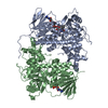
| ||||||||
|---|---|---|---|---|---|---|---|---|---|
| 1 |
| ||||||||
| Unit cell |
|
-Components
| #1: Protein | Mass: 62263.180 Da / Num. of mol.: 2 Source method: isolated from a genetically manipulated source Source: (gene. exp.) Streptomyces albogriseolus (bacteria) / Gene: thal, thdH / Plasmid: pET28a / Production host: Escherichia coli BL21(DE3) (bacteria) / References: UniProt: A1E280#2: Chemical | ChemComp-FAD / | Flavin adenine dinucleotide  Mass: 785.550 Da / Num. of mol.: 1 / Source method: obtained synthetically / Formula: C27H33N9O15P2 / Feature type: SUBJECT OF INVESTIGATION / Comment: FAD*YM Mass: 785.550 Da / Num. of mol.: 1 / Source method: obtained synthetically / Formula: C27H33N9O15P2 / Feature type: SUBJECT OF INVESTIGATION / Comment: FAD*YM#3: Chemical | ChemComp-AMP / | Adenosine monophosphate  Mass: 347.221 Da / Num. of mol.: 1 / Source method: obtained synthetically / Formula: C10H14N5O7P / Comment: AMP*YM Mass: 347.221 Da / Num. of mol.: 1 / Source method: obtained synthetically / Formula: C10H14N5O7P / Comment: AMP*YM#4: Water | ChemComp-HOH / | Water Mass: 18.015 Da / Num. of mol.: 731 / Source method: isolated from a natural source / Formula: H2O Mass: 18.015 Da / Num. of mol.: 731 / Source method: isolated from a natural source / Formula: H2OHas ligand of interest | Y | |
|---|
-Experimental details
-Experiment
| Experiment | Method: X-RAY DIFFRACTION / Number of used crystals: 1 |
|---|
- Sample preparation
| Crystal | Density Matthews: 2.14 Å3/Da / Density % sol: 42.66 % / Mosaicity: 0 ° |
|---|---|
| Crystal grow | Temperature: 293 K / Method: microbatch / pH: 7 / Details: 0.28-0.38M NaF, 23-31% w/v PEG 3350 |
-Data collection
| Diffraction | Mean temperature: 100 K / Serial crystal experiment: N |
|---|---|
| Diffraction source | Source: ROTATING ANODE / Type: BRUKER D8 QUEST / Wavelength: 1.54 Å |
| Detector | Type: BRUKER PHOTON 100 / Detector: CMOS / Date: Mar 22, 2017 |
| Radiation | Protocol: SINGLE WAVELENGTH / Monochromatic (M) / Laue (L): M / Scattering type: x-ray |
| Radiation wavelength | Wavelength: 1.54 Å / Relative weight: 1 |
| Reflection | Resolution: 1.91→20.64 Å / Num. obs: 77292 / % possible obs: 99 % / Redundancy: 7.1 % / CC1/2: 0.998 / Rmerge(I) obs: 0.075 / Rpim(I) all: 0.029 / Rrim(I) all: 0.081 / Net I/σ(I): 16.9 / Num. measured all: 547491 |
| Reflection shell | Resolution: 1.91→1.95 Å / Redundancy: 3.9 % / Rmerge(I) obs: 0.465 / Num. measured all: 15402 / Num. unique obs: 3948 / CC1/2: 0.834 / Rpim(I) all: 0.253 / Rrim(I) all: 0.533 / Net I/σ(I) obs: 3.3 / % possible all: 86.2 |
- Processing
| Software |
| |||||||||||||||||||||||||||||||||||||||||||||||||||||||||||||||||||||||||||||||||||||||||||||||||||||||||||||||||||||||||||||||||||||||||||||||||
|---|---|---|---|---|---|---|---|---|---|---|---|---|---|---|---|---|---|---|---|---|---|---|---|---|---|---|---|---|---|---|---|---|---|---|---|---|---|---|---|---|---|---|---|---|---|---|---|---|---|---|---|---|---|---|---|---|---|---|---|---|---|---|---|---|---|---|---|---|---|---|---|---|---|---|---|---|---|---|---|---|---|---|---|---|---|---|---|---|---|---|---|---|---|---|---|---|---|---|---|---|---|---|---|---|---|---|---|---|---|---|---|---|---|---|---|---|---|---|---|---|---|---|---|---|---|---|---|---|---|---|---|---|---|---|---|---|---|---|---|---|---|---|---|---|---|---|
| Refinement | Method to determine structure: MOLECULAR REPLACEMENT Starting model: 5HY5 Resolution: 1.91→20.64 Å / Cor.coef. Fo:Fc: 0.952 / Cor.coef. Fo:Fc free: 0.924 / SU B: 4.005 / SU ML: 0.115 / Cross valid method: THROUGHOUT / σ(F): 0 / ESU R: 0.171 / ESU R Free: 0.153 Details: HYDROGENS HAVE BEEN ADDED IN THE RIDING POSITIONS U VALUES : REFINED INDIVIDUALLY
| |||||||||||||||||||||||||||||||||||||||||||||||||||||||||||||||||||||||||||||||||||||||||||||||||||||||||||||||||||||||||||||||||||||||||||||||||
| Solvent computation | Ion probe radii: 0.8 Å / Shrinkage radii: 0.8 Å / VDW probe radii: 1.4 Å | |||||||||||||||||||||||||||||||||||||||||||||||||||||||||||||||||||||||||||||||||||||||||||||||||||||||||||||||||||||||||||||||||||||||||||||||||
| Displacement parameters | Biso max: 99.24 Å2 / Biso mean: 17.63 Å2 / Biso min: 5.15 Å2
| |||||||||||||||||||||||||||||||||||||||||||||||||||||||||||||||||||||||||||||||||||||||||||||||||||||||||||||||||||||||||||||||||||||||||||||||||
| Refinement step | Cycle: final / Resolution: 1.91→20.64 Å
| |||||||||||||||||||||||||||||||||||||||||||||||||||||||||||||||||||||||||||||||||||||||||||||||||||||||||||||||||||||||||||||||||||||||||||||||||
| Refine LS restraints |
| |||||||||||||||||||||||||||||||||||||||||||||||||||||||||||||||||||||||||||||||||||||||||||||||||||||||||||||||||||||||||||||||||||||||||||||||||
| LS refinement shell | Resolution: 1.91→1.96 Å / Rfactor Rfree error: 0 / Total num. of bins used: 20
|
