 Movie
Movie Controller

[English] 日本語
 Yorodumi
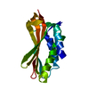
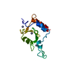
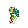
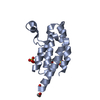
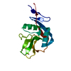
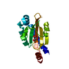
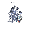
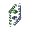
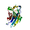
- PDB-7cqf: Crystal structure of PSD-95 PDZ3 fused with ADAM22 C-terminal peptide -
+ Open data

- Basic information
| Entry | Database: PDB / ID: 7cqf | ||||||
|---|---|---|---|---|---|---|---|
| Title | Crystal structure of PSD-95 PDZ3 fused with ADAM22 C-terminal peptide | ||||||
 Components Components | Disks large homolog 4,Disintegrin and metalloproteinase domain-containing protein 22 | ||||||
 Keywords Keywords |  SIGNALING PROTEIN / Scaffold protein / Membrane receptor SIGNALING PROTEIN / Scaffold protein / Membrane receptor | ||||||
| Function / homology |  Function and homology information Function and homology informationRHO GTPases activate CIT / LGI-ADAM interactions / positive regulation of AMPA glutamate receptor clustering / neuronal ion channel clustering / P2Y1 nucleotide receptor binding / Neurexins and neuroligins / beta-1 adrenergic receptor binding / neuroligin family protein binding / structural constituent of postsynaptic density / proximal dendrite ...RHO GTPases activate CIT / LGI-ADAM interactions / positive regulation of AMPA glutamate receptor clustering / neuronal ion channel clustering / P2Y1 nucleotide receptor binding / Neurexins and neuroligins / beta-1 adrenergic receptor binding / neuroligin family protein binding / structural constituent of postsynaptic density / proximal dendrite / receptor localization to synapse / positive regulation of neuron projection arborization / regulation of grooming behavior / synaptic vesicle maturation / cerebellar mossy fiber / cellular response to potassium ion / protein localization to synapse / vocalization behavior / LGI-ADAM interactions / Trafficking of AMPA receptors / neuron spine / dendritic branch / Activation of Ca-permeable Kainate Receptor / AMPA glutamate receptor clustering / juxtaparanode region of axon / establishment or maintenance of epithelial cell apical/basal polarity / dendritic spine morphogenesis / frizzled binding / negative regulation of cell adhesion / negative regulation of receptor internalization / postsynaptic neurotransmitter receptor diffusion trapping / neuron projection terminus / dendritic spine organization / acetylcholine receptor binding / positive regulation of synapse assembly / RAF/MAP kinase cascade / Synaptic adhesion-like molecules / neurotransmitter receptor localization to postsynaptic specialization membrane / positive regulation of dendrite morphogenesis / beta-2 adrenergic receptor binding / cortical cytoskeleton / regulation of neuronal synaptic plasticity / locomotory exploration behavior / regulation of NMDA receptor activity / social behavior / positive regulation of excitatory postsynaptic potential / kinesin binding / AMPA glutamate receptor complex / neuromuscular process controlling balance / excitatory synapse / D1 dopamine receptor binding / Unblocking of NMDA receptors, glutamate binding and activation / glutamate receptor binding / positive regulation of protein tyrosine kinase activity / positive regulation of synaptic transmission / ionotropic glutamate receptor binding / extrinsic component of cytoplasmic side of plasma membrane / dendrite cytoplasm / synaptic membrane / central nervous system development / PDZ domain binding / cell periphery / postsynaptic density membrane / regulation of long-term neuronal synaptic plasticity / neuromuscular junction / establishment of protein localization / cell-cell adhesion / metalloendopeptidase activity / cerebral cortex development / kinase binding / cell-cell junction / synaptic vesicle / integrin binding / cell junction / positive regulation of cytosolic calcium ion concentration / chemical synaptic transmission / postsynapse / scaffold protein binding / postsynaptic membrane / basolateral plasma membrane / protein phosphatase binding / protein-containing complex assembly / dendritic spine / postsynaptic density / cell adhesion / neuron projection / axon / signaling receptor binding / dendrite / synapse / glutamatergic synapse / protein-containing complex binding / protein kinase binding / endoplasmic reticulum / proteolysis / membrane / plasma membrane / cytosol / cytoplasmSimilarity search - Function | ||||||
| Biological species |  Rattus norvegicus (Norway rat) Rattus norvegicus (Norway rat) Homo sapiens (human) Homo sapiens (human) | ||||||
| Method | X-RAY DIFFRACTION / SYNCHROTRON / MOLECULAR REPLACEMENT / Resolution: 1.8 Å | ||||||
 Authors Authors | Yamagata, A. / Fukai, S. | ||||||
| Funding support |  Japan, 1items Japan, 1items
| ||||||
 Citation Citation | Journal: Proc.Natl.Acad.Sci.USA / Year: 2021 Title: LGI1-ADAM22-MAGUK configures transsynaptic nanoalignment for synaptic transmission and epilepsy prevention. Authors: Fukata, Y. / Chen, X. / Chiken, S. / Hirano, Y. / Yamagata, A. / Inahashi, H. / Sanbo, M. / Sano, H. / Goto, T. / Hirabayashi, M. / Kornau, H.C. / Pruss, H. / Nambu, A. / Fukai, S. / Nicoll, R.A. / Fukata, M. | ||||||
| History |
|
- Structure visualization
| Structure viewer | Molecule: MolmilJmol/JSmol |
|---|
- Downloads & links
-Download
| PDBx/mmCIF format | 7cqf.cif.gz | 40.8 KB | Display | PDBx/mmCIF format |
|---|---|---|---|---|
| PDB format | pdb7cqf.ent.gz | 25.4 KB | Display | PDB format |
| PDBx/mmJSON format | 7cqf.json.gz | Tree view | PDBx/mmJSON format | |
| Others |  Other downloads Other downloads |
-Validation report
| Arichive directory | https://data.pdbj.org/pub/pdb/validation_reports/cq/7cqfftp://data.pdbj.org/pub/pdb/validation_reports/cq/7cqf | HTTPS FTP |
|---|
-Related structure data
| Related structure data | 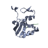 1tp3S S: Starting model for refinement |
|---|---|
| Similar structure data |

-Links
 PDBj
PDBj


- Assembly
| Deposited unit | 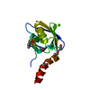
| ||||||||
|---|---|---|---|---|---|---|---|---|---|
| 1 |
| ||||||||
| Unit cell |
|
-Components
| #1: Protein | Mass: 16446.395 Da / Num. of mol.: 1 Source method: isolated from a genetically manipulated source Details: Fusion protein of PSD-95 PDZ3, linker, and ADAM22 C-terminal peptide Source: (gene. exp.) Rattus norvegicus (Norway rat), (gene. exp.) Homo sapiens (human)Gene: Dlg4, Dlgh4, Psd95, ADAM22, MDC2 / Production host:  Escherichia coli (E. coli) / References: UniProt: P31016, UniProt: Q9P0K1 Escherichia coli (E. coli) / References: UniProt: P31016, UniProt: Q9P0K1 | ||||
|---|---|---|---|---|---|
| #2: Chemical | ChemComp-CL / Chloride  Mass: 35.453 Da / Num. of mol.: 1 Mass: 35.453 Da / Num. of mol.: 1Source method: isolated from a genetically manipulated source Formula: Cl | ||||
| #3: Chemical | ChemComp-CHT / Choline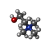  Mass: 104.171 Da / Num. of mol.: 1 / Source method: obtained synthetically / Formula: C5H14NO Mass: 104.171 Da / Num. of mol.: 1 / Source method: obtained synthetically / Formula: C5H14NO | ||||
| #4: Chemical | ChemComp-EDO / Ethylene glycol  Mass: 62.068 Da / Num. of mol.: 4 / Source method: obtained synthetically / Formula: C2H6O2 Mass: 62.068 Da / Num. of mol.: 4 / Source method: obtained synthetically / Formula: C2H6O2#5: Water | ChemComp-HOH / | Water Mass: 18.015 Da / Num. of mol.: 46 / Source method: isolated from a natural source / Formula: H2O Mass: 18.015 Da / Num. of mol.: 46 / Source method: isolated from a natural source / Formula: H2OHas ligand of interest | N | |
-Experimental details
-Experiment
| Experiment | Method: X-RAY DIFFRACTION / Number of used crystals: 1 |
|---|
- Sample preparation
| Crystal | Density Matthews: 1.75 Å3/Da / Density % sol: 29.79 % |
|---|---|
| Crystal grow | Temperature: 293 K / Method: vapor diffusion, sitting drop / pH: 7.5 Details: 0.2 M choline chloride, 0.1 M Tris-HCl (pH 7.5), 14 % (w/v) PEG 2000 MME |
-Data collection
| Diffraction | Mean temperature: 100 K / Serial crystal experiment: N |
|---|---|
| Diffraction source | Source: SYNCHROTRON / Site: SPring-8 / Beamline: BL41XU / Wavelength: 1 Å |
| Detector | Type: DECTRIS EIGER X 16M / Detector: PIXEL / Date: Apr 17, 2019 |
| Radiation | Protocol: SINGLE WAVELENGTH / Monochromatic (M) / Laue (L): M / Scattering type: x-ray |
| Radiation wavelength | Wavelength: 1 Å / Relative weight: 1 |
| Reflection | Resolution: 1.8→50 Å / Num. obs: 10624 / % possible obs: 100 % / Redundancy: 16.1 % / CC1/2: 0.999 / Rpim(I) all: 0.031 / Rrim(I) all: 0.134 / Rsym value: 0.13 / Net I/σ(I): 26.2 |
| Reflection shell | Resolution: 1.8→1.83 Å / Num. unique obs: 532 / CC1/2: 0.944 / Rpim(I) all: 0.262 / Rrim(I) all: 0.791 / Rsym value: 0.745 |
- Processing
| Software |
| ||||||||||||||||||||||||||||||||||||||||||||||||||||||||||||||||||||||||||||||||||||||||||||||||||||||||||||||||||||||||||||||||||||||||||||||||||||||||||||||||||||||||||||||||||||||
|---|---|---|---|---|---|---|---|---|---|---|---|---|---|---|---|---|---|---|---|---|---|---|---|---|---|---|---|---|---|---|---|---|---|---|---|---|---|---|---|---|---|---|---|---|---|---|---|---|---|---|---|---|---|---|---|---|---|---|---|---|---|---|---|---|---|---|---|---|---|---|---|---|---|---|---|---|---|---|---|---|---|---|---|---|---|---|---|---|---|---|---|---|---|---|---|---|---|---|---|---|---|---|---|---|---|---|---|---|---|---|---|---|---|---|---|---|---|---|---|---|---|---|---|---|---|---|---|---|---|---|---|---|---|---|---|---|---|---|---|---|---|---|---|---|---|---|---|---|---|---|---|---|---|---|---|---|---|---|---|---|---|---|---|---|---|---|---|---|---|---|---|---|---|---|---|---|---|---|---|---|---|---|---|
| Refinement | Method to determine structure: MOLECULAR REPLACEMENT Starting model: 1TP3 Resolution: 1.8→36.64 Å / Cor.coef. Fo:Fc: 0.966 / Cor.coef. Fo:Fc free: 0.941 / SU B: 2.781 / SU ML: 0.084 / Cross valid method: THROUGHOUT / ESU R: 0.12 / ESU R Free: 0.116 / Stereochemistry target values: MAXIMUM LIKELIHOOD / Details: HYDROGENS HAVE BEEN ADDED IN THE RIDING POSITIONS
| ||||||||||||||||||||||||||||||||||||||||||||||||||||||||||||||||||||||||||||||||||||||||||||||||||||||||||||||||||||||||||||||||||||||||||||||||||||||||||||||||||||||||||||||||||||||
| Solvent computation | Ion probe radii: 0.8 Å / Shrinkage radii: 0.8 Å / VDW probe radii: 1.2 Å / Solvent model: MASK | ||||||||||||||||||||||||||||||||||||||||||||||||||||||||||||||||||||||||||||||||||||||||||||||||||||||||||||||||||||||||||||||||||||||||||||||||||||||||||||||||||||||||||||||||||||||
| Displacement parameters | Biso mean: 31.53 Å2
| ||||||||||||||||||||||||||||||||||||||||||||||||||||||||||||||||||||||||||||||||||||||||||||||||||||||||||||||||||||||||||||||||||||||||||||||||||||||||||||||||||||||||||||||||||||||
| Refinement step | Cycle: 1 / Resolution: 1.8→36.64 Å
| ||||||||||||||||||||||||||||||||||||||||||||||||||||||||||||||||||||||||||||||||||||||||||||||||||||||||||||||||||||||||||||||||||||||||||||||||||||||||||||||||||||||||||||||||||||||
| Refine LS restraints |
|