 Movie
Movie Controller

[English] 日本語
 Yorodumi
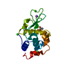
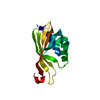
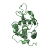
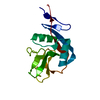
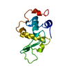
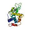
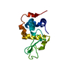
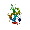
- PDB-1zv1: Crystal structure of the dimerization domain of doublesex protein... -
+ Open data

- Basic information
| Entry | Database: PDB / ID: 1zv1 | ||||||
|---|---|---|---|---|---|---|---|
| Title | Crystal structure of the dimerization domain of doublesex protein from D. melanogaster | ||||||
 Components Components | Doublesex protein | ||||||
 Keywords Keywords |  PROTEIN BINDING / doublesex / uba domain / dimerization / sex determination / transcription factor PROTEIN BINDING / doublesex / uba domain / dimerization / sex determination / transcription factor | ||||||
| Function / homology |  Function and homology information Function and homology informationimaginal disc-derived male genitalia development / male analia development / female analia development / female sex differentiation / sex-specific pigmentation / female somatic sex determination / imaginal disc-derived female genitalia development / sex comb development / negative regulation of developmental pigmentation / somatic sex determination ...imaginal disc-derived male genitalia development / male analia development / female analia development / female sex differentiation / sex-specific pigmentation / female somatic sex determination / imaginal disc-derived female genitalia development / sex comb development / negative regulation of developmental pigmentation / somatic sex determination / genital disc development / male sex differentiation / courtship behavior / male courtship behavior, veined wing generated song production / sex determination / male courtship behavior / sex differentiation / axon midline choice point recognition / RNA polymerase II transcription regulatory region sequence-specific DNA binding / DNA-binding transcription repressor activity, RNA polymerase II-specific / DNA-binding transcription activator activity, RNA polymerase II-specific / DNA-binding transcription factor activity, RNA polymerase II-specific / RNA polymerase II cis-regulatory region sequence-specific DNA binding / negative regulation of DNA-templated transcription / regulation of transcription by RNA polymerase II / positive regulation of DNA-templated transcription / protein homodimerization activity / positive regulation of transcription by RNA polymerase II / zinc ion binding / nucleusSimilarity search - Function | ||||||
| Biological species |  Drosophila melanogaster (fruit fly) Drosophila melanogaster (fruit fly) | ||||||
| Method | X-RAY DIFFRACTION / SYNCHROTRON / SAD / Resolution: 1.6 Å | ||||||
 Authors Authors | Weiss, M.A. / Bayrer, J.R. / Wan, Z. / Li, B. / Phillips, N.B. | ||||||
 Citation Citation | Journal: J.Biol.Chem. / Year: 2005 Title: Dimerization of doublesex is mediated by a cryptic ubiquitin-associated domain fold: implications for sex-specific gene regulation Authors: Bayrer, J.R. / Zhang, W. / Weiss, M.A. | ||||||
| History |
|
- Structure visualization
| Structure viewer | Molecule: MolmilJmol/JSmol |
|---|
- Downloads & links
-Download
| PDBx/mmCIF format | 1zv1.cif.gz | 39.1 KB | Display | PDBx/mmCIF format |
|---|---|---|---|---|
| PDB format | pdb1zv1.ent.gz | 27.6 KB | Display | PDB format |
| PDBx/mmJSON format | 1zv1.json.gz | Tree view | PDBx/mmJSON format | |
| Others |  Other downloads Other downloads |
-Validation report
| Arichive directory | https://data.pdbj.org/pub/pdb/validation_reports/zv/1zv1ftp://data.pdbj.org/pub/pdb/validation_reports/zv/1zv1 | HTTPS FTP |
|---|
-Related structure data
| Similar structure data |
|---|


-Links
 PDBj
PDBj- Assembly
| Deposited unit | 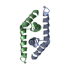
| ||||||||
|---|---|---|---|---|---|---|---|---|---|
| 1 |
| ||||||||
| Unit cell |
| ||||||||
| Details | The biological assembly is a dimer found entirely in the asymmetric unit. |
-Components
| #1: Protein | Mass: 7745.735 Da / Num. of mol.: 2 / Fragment: DIMERIZATION DOMAIN Source method: isolated from a genetically manipulated source Source: (gene. exp.) Drosophila melanogaster (fruit fly) / Gene: dsx / Production host:  Escherichia coli (E. coli) / References: UniProt: P23023 Escherichia coli (E. coli) / References: UniProt: P23023#2: Water | ChemComp-HOH / | Water Mass: 18.015 Da / Num. of mol.: 126 / Source method: isolated from a natural source / Formula: H2O Mass: 18.015 Da / Num. of mol.: 126 / Source method: isolated from a natural source / Formula: H2O |
|---|
-Experimental details
-Experiment
| Experiment | Method: X-RAY DIFFRACTION / Number of used crystals: 2 |
|---|
- Sample preparation
| Crystal | Density Matthews: 1.787 Å3/Da / Density % sol: 28.53 % |
|---|---|
| Crystal grow | Temperature: 298 K / Method: vapor diffusion, hanging drop / pH: 7.4 Details: ammonium sulfate, isopropanol, tris, sodium chloride , pH 7.4, VAPOR DIFFUSION, HANGING DROP, temperature 298K |
-Data collection
| Diffraction |
| ||||||||||||||||||
|---|---|---|---|---|---|---|---|---|---|---|---|---|---|---|---|---|---|---|---|
| Diffraction source |
| ||||||||||||||||||
| Detector |
| ||||||||||||||||||
| Radiation |
| ||||||||||||||||||
| Radiation wavelength |
| ||||||||||||||||||
| Reflection | Resolution: 1.6→20 Å / Num. all: 15159 / Num. obs: 15159 / Observed criterion σ(F): 0 / Observed criterion σ(I): -3 / Redundancy: 10.9 % / Rmerge(I) obs: 0.038 / Net I/σ(I): 62.3 | ||||||||||||||||||
| Reflection shell | Resolution: 1.6→1.66 Å / Rmerge(I) obs: 0.182 / Mean I/σ(I) obs: 11 / % possible all: 96.9 |

- Processing
| Software |
| ||||||||||||||||||||||||||||||||||||||||||||||||||||||||||||||||||||||||||||||||||||||||||||||||||||||||||||||||||||||||||||||||||||||||||||||||||||||||||||||||||||||||||
|---|---|---|---|---|---|---|---|---|---|---|---|---|---|---|---|---|---|---|---|---|---|---|---|---|---|---|---|---|---|---|---|---|---|---|---|---|---|---|---|---|---|---|---|---|---|---|---|---|---|---|---|---|---|---|---|---|---|---|---|---|---|---|---|---|---|---|---|---|---|---|---|---|---|---|---|---|---|---|---|---|---|---|---|---|---|---|---|---|---|---|---|---|---|---|---|---|---|---|---|---|---|---|---|---|---|---|---|---|---|---|---|---|---|---|---|---|---|---|---|---|---|---|---|---|---|---|---|---|---|---|---|---|---|---|---|---|---|---|---|---|---|---|---|---|---|---|---|---|---|---|---|---|---|---|---|---|---|---|---|---|---|---|---|---|---|---|---|---|---|---|---|
| Refinement | Method to determine structure: SAD / Resolution: 1.6→10.86 Å / Cor.coef. Fo:Fc: 0.947 / Cor.coef. Fo:Fc free: 0.925 / SU B: 1.707 / SU ML: 0.063 / Isotropic thermal model: RESTRAINED / Cross valid method: THROUGHOUT / ESU R: 0.115 / ESU R Free: 0.118 Stereochemistry target values: MAXIMUM LIKELIHOOD WITH PHASES Details: HYDROGENS HAVE BEEN ADDED IN THE RIDING POSITIONS
| ||||||||||||||||||||||||||||||||||||||||||||||||||||||||||||||||||||||||||||||||||||||||||||||||||||||||||||||||||||||||||||||||||||||||||||||||||||||||||||||||||||||||||
| Solvent computation | Ion probe radii: 0.8 Å / Shrinkage radii: 0.8 Å / VDW probe radii: 1.2 Å / Solvent model: MASK | ||||||||||||||||||||||||||||||||||||||||||||||||||||||||||||||||||||||||||||||||||||||||||||||||||||||||||||||||||||||||||||||||||||||||||||||||||||||||||||||||||||||||||
| Displacement parameters | Biso mean: 27.969 Å2
| ||||||||||||||||||||||||||||||||||||||||||||||||||||||||||||||||||||||||||||||||||||||||||||||||||||||||||||||||||||||||||||||||||||||||||||||||||||||||||||||||||||||||||
| Refine analyze |
| ||||||||||||||||||||||||||||||||||||||||||||||||||||||||||||||||||||||||||||||||||||||||||||||||||||||||||||||||||||||||||||||||||||||||||||||||||||||||||||||||||||||||||
| Refinement step | Cycle: LAST / Resolution: 1.6→10.86 Å
| ||||||||||||||||||||||||||||||||||||||||||||||||||||||||||||||||||||||||||||||||||||||||||||||||||||||||||||||||||||||||||||||||||||||||||||||||||||||||||||||||||||||||||
| Refine LS restraints |
| ||||||||||||||||||||||||||||||||||||||||||||||||||||||||||||||||||||||||||||||||||||||||||||||||||||||||||||||||||||||||||||||||||||||||||||||||||||||||||||||||||||||||||
| LS refinement shell | Resolution: 1.6→1.641 Å / Total num. of bins used: 20
| ||||||||||||||||||||||||||||||||||||||||||||||||||||||||||||||||||||||||||||||||||||||||||||||||||||||||||||||||||||||||||||||||||||||||||||||||||||||||||||||||||||||||||
| Xplor file |
|
