 Movie
Movie Controller

[English] 日本語
 Yorodumi
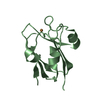
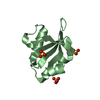
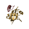
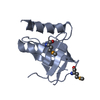
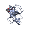
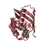
- PDB-7clt: Crystal structure of the EFhd1/Swiprosin-2, a mitochondrial actin... -
+ Open data

- Basic information
| Entry | Database: PDB / ID: 7clt | ||||||
|---|---|---|---|---|---|---|---|
| Title | Crystal structure of the EFhd1/Swiprosin-2, a mitochondrial actin-binding protein | ||||||
 Components Components | EF-hand domain-containing protein D1 | ||||||
 Keywords Keywords | METAL BINDING PROTEIN /  mitochondria / calcium-binding mitochondria / calcium-binding | ||||||
| Function / homology |  Function and homology information Function and homology informationregulation of cellular hyperosmotic salinity response / calcium ion sensor activity / neuron projection development / mitochondrial inner membrane / calcium ion binding / mitochondrionSimilarity search - Function | ||||||
| Biological species |  Mus musculus (house mouse) Mus musculus (house mouse) | ||||||
| Method | X-RAY DIFFRACTION / SYNCHROTRON / MOLECULAR REPLACEMENT / Resolution: 2.07380981063 Å | ||||||
 Authors Authors | Mun, S.A. / Park, J. / Park, K.R. / Lee, Y. / Kang, J.Y. / Park, T. / Jin, M. / Yang, J. / Jun, C.D. / Eom, S.H. | ||||||
| Funding support |  Korea, Republic Of, 1items Korea, Republic Of, 1items
| ||||||
 Citation Citation | Journal: Front Cell Dev Biol / Year: 2020 Title: Structural and Biochemical Characterization of EFhd1/Swiprosin-2, an Actin-Binding Protein in Mitochondria. Authors: Mun, S.A. / Park, J. / Park, K.R. / Lee, Y. / Kang, J.Y. / Park, T. / Jin, M. / Yang, J. / Jun, C.D. / Eom, S.H. | ||||||
| History |
|
- Structure visualization
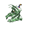
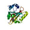
| Structure viewer | Molecule: MolmilJmol/JSmol |
|---|
- Downloads & links
-Download
| PDBx/mmCIF format | 7clt.cif.gz | 43.7 KB | Display | PDBx/mmCIF format |
|---|---|---|---|---|
| PDB format | pdb7clt.ent.gz | 23 KB | Display | PDB format |
| PDBx/mmJSON format | 7clt.json.gz | Tree view | PDBx/mmJSON format | |
| Others |  Other downloads Other downloads |
-Validation report
| Arichive directory | https://data.pdbj.org/pub/pdb/validation_reports/cl/7cltftp://data.pdbj.org/pub/pdb/validation_reports/cl/7clt | HTTPS FTP |
|---|
-Related structure data
| Related structure data |  5i2lS S: Starting model for refinement |
|---|---|
| Similar structure data |

-Links
 PDBj
PDBj- Assembly
| Deposited unit | 
| ||||||||||||
|---|---|---|---|---|---|---|---|---|---|---|---|---|---|
| 1 |
| ||||||||||||
| Unit cell |
|
-Components
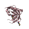
| #1: Protein | Mass: 15192.491 Da / Num. of mol.: 1 Source method: isolated from a genetically manipulated source Source: (gene. exp.) Mus musculus (house mouse) / Gene: Efhd1, Sws2 / Production host:  Escherichia coli BL21(DE3) (bacteria) / Strain (production host): BL21(DE3) / References: UniProt: Q9D4J1 Escherichia coli BL21(DE3) (bacteria) / Strain (production host): BL21(DE3) / References: UniProt: Q9D4J1 | ||||||||
|---|---|---|---|---|---|---|---|---|---|
| #2: Chemical |   Mass: 40.078 Da / Num. of mol.: 2 / Source method: obtained synthetically / Formula: Ca / Feature type: SUBJECT OF INVESTIGATION Mass: 40.078 Da / Num. of mol.: 2 / Source method: obtained synthetically / Formula: Ca / Feature type: SUBJECT OF INVESTIGATION#3: Chemical |   Mass: 65.409 Da / Num. of mol.: 2 / Source method: obtained synthetically / Formula: Zn / Feature type: SUBJECT OF INVESTIGATION Mass: 65.409 Da / Num. of mol.: 2 / Source method: obtained synthetically / Formula: Zn / Feature type: SUBJECT OF INVESTIGATION#4: Chemical | ChemComp-GOL / | Glycerol  Mass: 92.094 Da / Num. of mol.: 1 / Source method: obtained synthetically / Formula: C3H8O3 / Feature type: SUBJECT OF INVESTIGATION Mass: 92.094 Da / Num. of mol.: 1 / Source method: obtained synthetically / Formula: C3H8O3 / Feature type: SUBJECT OF INVESTIGATION#5: Water | ChemComp-HOH / | Water Mass: 18.015 Da / Num. of mol.: 23 / Source method: isolated from a natural source / Formula: H2O Mass: 18.015 Da / Num. of mol.: 23 / Source method: isolated from a natural source / Formula: H2OHas ligand of interest | Y | |
-Experimental details
-Experiment
| Experiment | Method: X-RAY DIFFRACTION / Number of used crystals: 1 |
|---|
- Sample preparation
| Crystal | Density Matthews: 2.15 Å3/Da / Density % sol: 42.9 % |
|---|---|
| Crystal grow | Temperature: 293 K / Method: vapor diffusion, sitting drop / pH: 7.5 Details: 100 mM HEPES-NaOH, 5 mM ZnSO4, 25% (v/v) Jeffamine ED-2001 |
-Data collection
| Diffraction | Mean temperature: 100 K / Serial crystal experiment: N |
|---|---|
| Diffraction source | Source: SYNCHROTRON / Site: PAL/PLS / Beamline: 5C (4A) / Wavelength: 0.9794 Å |
| Detector | Type: DECTRIS PILATUS 6M / Detector: PIXEL / Date: Mar 30, 2016 |
| Radiation | Protocol: SINGLE WAVELENGTH / Monochromatic (M) / Laue (L): M / Scattering type: x-ray |
| Radiation wavelength | Wavelength: 0.9794 Å / Relative weight: 1 |
| Reflection | Resolution: 2.07→50 Å / Num. obs: 8434 / % possible obs: 99.2 % / Redundancy: 4.9 % / Biso Wilson estimate: 24 Å2 / Rmerge(I) obs: 0.046 / Net I/σ(I): 11.1 |
| Reflection shell | Resolution: 2.07→2.11 Å / Rmerge(I) obs: 0.59 / Num. unique obs: 403 |
- Processing
| Software |
| ||||||||||||||||||||||||||||
|---|---|---|---|---|---|---|---|---|---|---|---|---|---|---|---|---|---|---|---|---|---|---|---|---|---|---|---|---|---|
| Refinement | Method to determine structure: MOLECULAR REPLACEMENT Starting model: 5I2L Resolution: 2.07380981063→43.5995 Å / SU ML: 0.215981547602 / Cross valid method: FREE R-VALUE / σ(F): 1.36030208239 / Phase error: 22.4306799448 Stereochemistry target values: GeoStd + Monomer Library + CDL v1.2
| ||||||||||||||||||||||||||||
| Solvent computation | Shrinkage radii: 0.9 Å / VDW probe radii: 1.11 Å / Solvent model: FLAT BULK SOLVENT MODEL | ||||||||||||||||||||||||||||
| Displacement parameters | Biso mean: 31.6235678918 Å2 | ||||||||||||||||||||||||||||
| Refinement step | Cycle: LAST / Resolution: 2.07380981063→43.5995 Å
| ||||||||||||||||||||||||||||
| Refine LS restraints |
| ||||||||||||||||||||||||||||
| LS refinement shell |
|
