 Movie
Movie Controller


+ Open data

- Basic information
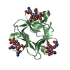
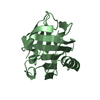
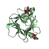
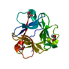
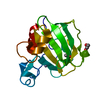
| Entry | Database: PDB / ID: 7ckv | ||||||
|---|---|---|---|---|---|---|---|
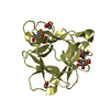
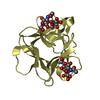
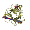
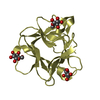
| Title | Crystal structure of Cyanobacteriochrome GAF domain in Pr state | ||||||
 Components Components | RcaE | ||||||
 Keywords Keywords |  SIGNALING PROTEIN / Cyanobacteriochrome SIGNALING PROTEIN / Cyanobacteriochrome | ||||||
| Function / homology |  Function and homology information Function and homology information | ||||||
| Biological species |  Microchaete diplosiphon (bacteria) Microchaete diplosiphon (bacteria) | ||||||
| Method | X-RAY DIFFRACTION / SYNCHROTRON / MOLECULAR REPLACEMENT / Resolution: 1.63 Å | ||||||
 Authors Authors | Nagae, T. / Koizumi, T. / Hirose, Y. / Mishima, M. | ||||||
 Citation Citation | Journal: Proc.Natl.Acad.Sci.USA / Year: 2021 Title: Structural basis of the protochromic green/red photocycle of the chromatic acclimation sensor RcaE. Authors: Nagae, T. / Unno, M. / Koizumi, T. / Miyanoiri, Y. / Fujisawa, T. / Masui, K. / Kamo, T. / Wada, K. / Eki, T. / Ito, Y. / Hirose, Y. / Mishima, M. | ||||||
| History |
|
- Structure visualization
| Structure viewer | Molecule: MolmilJmol/JSmol |
|---|
- Downloads & links
-Download
| PDBx/mmCIF format | 7ckv.cif.gz | 87.2 KB | Display | PDBx/mmCIF format |
|---|---|---|---|---|
| PDB format | pdb7ckv.ent.gz | 61 KB | Display | PDB format |
| PDBx/mmJSON format | 7ckv.json.gz | Tree view | PDBx/mmJSON format | |
| Others |  Other downloads Other downloads |
-Validation report
| Arichive directory | https://data.pdbj.org/pub/pdb/validation_reports/ck/7ckvftp://data.pdbj.org/pub/pdb/validation_reports/ck/7ckv | HTTPS FTP |
|---|
-Related structure data
| Related structure data | 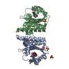 3vv4S S: Starting model for refinement |
|---|---|
| Similar structure data |

-Links
 PDBj
PDBj


- Assembly
| Deposited unit | 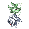
| ||||||||
|---|---|---|---|---|---|---|---|---|---|
| 1 | 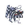
| ||||||||
| 2 | 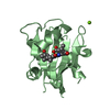
| ||||||||
| Unit cell |
|
-Components
| #1: Protein | Mass: 20391.125 Da / Num. of mol.: 2 / Fragment: GAF domain Source method: isolated from a genetically manipulated source Source: (gene. exp.) Microchaete diplosiphon (bacteria) / Gene: rcaE / Production host: Escherichia coli BL21 (bacteria) / References: UniProt: Q47897#2: Chemical | Phycocyanobilin  Mass: 588.694 Da / Num. of mol.: 2 / Source method: obtained synthetically / Formula: C33H40N4O6 / Feature type: SUBJECT OF INVESTIGATION Mass: 588.694 Da / Num. of mol.: 2 / Source method: obtained synthetically / Formula: C33H40N4O6 / Feature type: SUBJECT OF INVESTIGATION#3: Chemical | ChemComp-MG / |   Mass: 24.305 Da / Num. of mol.: 1 / Source method: obtained synthetically / Formula: Mg Mass: 24.305 Da / Num. of mol.: 1 / Source method: obtained synthetically / Formula: Mg#4: Water | ChemComp-HOH / | Water Mass: 18.015 Da / Num. of mol.: 346 / Source method: isolated from a natural source / Formula: H2O Mass: 18.015 Da / Num. of mol.: 346 / Source method: isolated from a natural source / Formula: H2OHas ligand of interest | Y | |
|---|
-Experimental details
-Experiment
| Experiment | Method: X-RAY DIFFRACTION / Number of used crystals: 1 |
|---|
- Sample preparation
| Crystal | Density Matthews: 1.86 Å3/Da / Density % sol: 33.69 % |
|---|---|
| Crystal grow | Temperature: 293 K / Method: vapor diffusion, hanging drop / pH: 7.2 / Details: 27% PEG 4000, 0.2M MgCl2, 0.1M Tris-HCl |
-Data collection
| Diffraction | Mean temperature: 95 K / Serial crystal experiment: N |
|---|---|
| Diffraction source | Source: SYNCHROTRON / Site: AichiSR  / Beamline: BL2S1 / Wavelength: 1.12 Å / Beamline: BL2S1 / Wavelength: 1.12 Å |
| Detector | Type: ADSC QUANTUM 270 / Detector: CCD / Date: Jan 29, 2020 |
| Radiation | Monochromator: Ge(111) / Protocol: SINGLE WAVELENGTH / Monochromatic (M) / Laue (L): M / Scattering type: x-ray |
| Radiation wavelength | Wavelength: 1.12 Å / Relative weight: 1 |
| Reflection | Resolution: 1.63→41.79 Å / Num. obs: 36876 / % possible obs: 99.6 % / Redundancy: 3.9 % / Biso Wilson estimate: 15 Å2 / CC1/2: 0.996 / Rmerge(I) obs: 0.046 / Rpim(I) all: 0.027 / Rrim(I) all: 0.054 / Χ2: 0.57 / Net I/σ(I): 13.3 |
| Reflection shell | Resolution: 1.63→1.66 Å / Redundancy: 3.9 % / Rmerge(I) obs: 0.483 / Mean I/σ(I) obs: 2.2 / Num. unique obs: 1810 / CC1/2: 0.837 / Rpim(I) all: 0.285 / Rrim(I) all: 0.562 / Χ2: 0.57 / % possible all: 99.9 |
- Processing
| Software |
| |||||||||||||||||||||||||||||||||||||||||||||||||||||||||||||||||||||||||||||||||||||||||||||||||||||||||||||||||||||||||||||||||||||||||||||||||||||||||||
|---|---|---|---|---|---|---|---|---|---|---|---|---|---|---|---|---|---|---|---|---|---|---|---|---|---|---|---|---|---|---|---|---|---|---|---|---|---|---|---|---|---|---|---|---|---|---|---|---|---|---|---|---|---|---|---|---|---|---|---|---|---|---|---|---|---|---|---|---|---|---|---|---|---|---|---|---|---|---|---|---|---|---|---|---|---|---|---|---|---|---|---|---|---|---|---|---|---|---|---|---|---|---|---|---|---|---|---|---|---|---|---|---|---|---|---|---|---|---|---|---|---|---|---|---|---|---|---|---|---|---|---|---|---|---|---|---|---|---|---|---|---|---|---|---|---|---|---|---|---|---|---|---|---|---|---|---|
| Refinement | Method to determine structure: MOLECULAR REPLACEMENT Starting model: 3vv4 Resolution: 1.63→38.663 Å / Cor.coef. Fo:Fc: 0.97 / Cor.coef. Fo:Fc free: 0.957 / SU B: 1.973 / SU ML: 0.066 / Cross valid method: FREE R-VALUE / ESU R: 0.089 / ESU R Free: 0.093 Details: Hydrogens have been added in their riding positions
| |||||||||||||||||||||||||||||||||||||||||||||||||||||||||||||||||||||||||||||||||||||||||||||||||||||||||||||||||||||||||||||||||||||||||||||||||||||||||||
| Solvent computation | Ion probe radii: 0.8 Å / Shrinkage radii: 0.8 Å / VDW probe radii: 1.2 Å / Solvent model: MASK BULK SOLVENT | |||||||||||||||||||||||||||||||||||||||||||||||||||||||||||||||||||||||||||||||||||||||||||||||||||||||||||||||||||||||||||||||||||||||||||||||||||||||||||
| Displacement parameters | Biso mean: 20.225 Å2
| |||||||||||||||||||||||||||||||||||||||||||||||||||||||||||||||||||||||||||||||||||||||||||||||||||||||||||||||||||||||||||||||||||||||||||||||||||||||||||
| Refinement step | Cycle: LAST / Resolution: 1.63→38.663 Å
| |||||||||||||||||||||||||||||||||||||||||||||||||||||||||||||||||||||||||||||||||||||||||||||||||||||||||||||||||||||||||||||||||||||||||||||||||||||||||||
| Refine LS restraints |
| |||||||||||||||||||||||||||||||||||||||||||||||||||||||||||||||||||||||||||||||||||||||||||||||||||||||||||||||||||||||||||||||||||||||||||||||||||||||||||
| LS refinement shell |
|
