 Movie
Movie Controller


+ Open data

- Basic information
| Entry | Database: PDB / ID: 3vv4 | ||||||
|---|---|---|---|---|---|---|---|
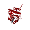
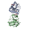
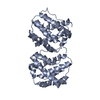
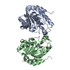
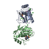
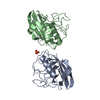
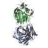
| Title | Crystal structure of cyanobacteriochrome TePixJ GAF domain | ||||||
 Components Components | Methyl-accepting chemotaxis protein Methyl-accepting chemotaxis proteins Methyl-accepting chemotaxis proteins | ||||||
 Keywords Keywords | SIGNALING PROTEIN / cyanobacteriochrome / phycoviolobilin binding | ||||||
| Function / homology |  Function and homology informationchemotaxis / lysozyme activity / signal transduction / metal ion binding / plasma membrane Function and homology informationchemotaxis / lysozyme activity / signal transduction / metal ion binding / plasma membraneSimilarity search - Function | ||||||
| Biological species |   Thermosynechococcus elongatus (bacteria) Thermosynechococcus elongatus (bacteria) | ||||||
| Method | X-RAY DIFFRACTION / SYNCHROTRON / MOLECULAR REPLACEMENT / Resolution: 2 Å | ||||||
 Authors Authors | Ishizuka, T. / Narikawa, R. / Muraki, N. / Shiba, T. / Kurisu, G. / Ikeuchi, M. | ||||||
 Citation Citation | Journal: Proc.Natl.Acad.Sci.USA / Year: 2013 Title: Structures of cyanobacteriochromes from phototaxis regulators AnPixJ and TePixJ reveal general and specific photoconversion mechanism Authors: Narikawa, R. / Ishizuka, T. / Muraki, N. / Shiba, T. / Kurisu, G. / Ikeuchi, M. | ||||||
| History |
|
- Structure visualization
| Structure viewer | Molecule: MolmilJmol/JSmol |
|---|
- Downloads & links
-Download
| PDBx/mmCIF format | 3vv4.cif.gz | 141.7 KB | Display | PDBx/mmCIF format |
|---|---|---|---|---|
| PDB format | pdb3vv4.ent.gz | 118.7 KB | Display | PDB format |
| PDBx/mmJSON format | 3vv4.json.gz | Tree view | PDBx/mmJSON format | |
| Others |  Other downloads Other downloads |
-Validation report
| Arichive directory | https://data.pdbj.org/pub/pdb/validation_reports/vv/3vv4ftp://data.pdbj.org/pub/pdb/validation_reports/vv/3vv4 | HTTPS FTP |
|---|
-Related structure data


-Links
 PDBj
PDBj


- Assembly
| Deposited unit | 
| |||||||||||||||||||||||||||
|---|---|---|---|---|---|---|---|---|---|---|---|---|---|---|---|---|---|---|---|---|---|---|---|---|---|---|---|---|
| 1 | 
| |||||||||||||||||||||||||||
| 2 | 
| |||||||||||||||||||||||||||
| Unit cell |
| |||||||||||||||||||||||||||
| Noncrystallographic symmetry (NCS) | NCS domain:
NCS domain segments:
|
-Components
| #1: Protein | Methyl-accepting chemotaxis proteins / TePixJ Mass: 22291.098 Da / Num. of mol.: 2 / Fragment: GAF domain, UNP residues 430-591 Source method: isolated from a genetically manipulated source Source: (gene. exp.) Thermosynechococcus elongatus (bacteria)Strain: BP-1 / Gene: tll0569 / Production host: Synechocystis (bacteria) / Strain (production host): PCC6803 / References: UniProt: Q8DLC7#2: Chemical |   Mass: 588.694 Da / Num. of mol.: 2 / Source method: obtained synthetically / Formula: C33H40N4O6 Mass: 588.694 Da / Num. of mol.: 2 / Source method: obtained synthetically / Formula: C33H40N4O6#3: Chemical | ChemComp-SO4 / Sulfate  Mass: 96.063 Da / Num. of mol.: 4 / Source method: obtained synthetically / Formula: SO4 Mass: 96.063 Da / Num. of mol.: 4 / Source method: obtained synthetically / Formula: SO4#4: Water | ChemComp-HOH / | Water Mass: 18.015 Da / Num. of mol.: 111 / Source method: isolated from a natural source / Formula: H2O Mass: 18.015 Da / Num. of mol.: 111 / Source method: isolated from a natural source / Formula: H2O |
|---|
-Experimental details
-Experiment
| Experiment | Method: X-RAY DIFFRACTION / Number of used crystals: 1 |
|---|
- Sample preparation
| Crystal | Density Matthews: 2.87 Å3/Da / Density % sol: 57.07 % |
|---|---|
| Crystal grow | Temperature: 277 K / Method: vapor diffusion, hanging drop / pH: 5.8 Details: 1.15M ammonium sulfate, 100mM MES buffer, pH 5.8, VAPOR DIFFUSION, HANGING DROP, temperature 277K |
-Data collection
| Diffraction | Mean temperature: 100 K |
|---|---|
| Diffraction source | Source: SYNCHROTRON / Site: Photon Factory  / Beamline: BL-17A / Wavelength: 1 Å / Beamline: BL-17A / Wavelength: 1 Å |
| Detector | Type: ADSC QUANTUM 270 / Detector: CCD / Date: Nov 11, 2011 |
| Radiation | Monochromator: Si 111 / Protocol: SINGLE WAVELENGTH / Monochromatic (M) / Laue (L): M / Scattering type: x-ray |
| Radiation wavelength | Wavelength: 1 Å / Relative weight: 1 |
| Reflection | Resolution: 1.95→100 Å / Num. all: 39334 / Num. obs: 36534 / % possible obs: 92.88 % / Observed criterion σ(F): 0 / Observed criterion σ(I): 2 |
| Reflection shell | Resolution: 1.95→2.02 Å / Rmerge(I) obs: 0.566 / Mean I/σ(I) obs: 2.2 / % possible all: 71 |
- Processing
| Software |
| |||||||||||||||||||||||||||||||||||||||||||||||||||||||||||||||||||||||||||
|---|---|---|---|---|---|---|---|---|---|---|---|---|---|---|---|---|---|---|---|---|---|---|---|---|---|---|---|---|---|---|---|---|---|---|---|---|---|---|---|---|---|---|---|---|---|---|---|---|---|---|---|---|---|---|---|---|---|---|---|---|---|---|---|---|---|---|---|---|---|---|---|---|---|---|---|---|
| Refinement | Method to determine structure: MOLECULAR REPLACEMENT / Resolution: 2→59.02 Å / Cor.coef. Fo:Fc: 0.963 / Cor.coef. Fo:Fc free: 0.95 / Occupancy max: 1 / Occupancy min: 0.5 / SU B: 6.994 / SU ML: 0.097 / Cross valid method: THROUGHOUT / σ(F): 0 / ESU R: 0.144 / ESU R Free: 0.139 / Stereochemistry target values: MAXIMUM LIKELIHOOD Details: HYDROGENS HAVE BEEN USED IF PRESENT IN THE INPUT U VALUES: RESIDUAL ONLY
| |||||||||||||||||||||||||||||||||||||||||||||||||||||||||||||||||||||||||||
| Solvent computation | Ion probe radii: 0.8 Å / Shrinkage radii: 0.8 Å / VDW probe radii: 1.2 Å / Solvent model: MASK | |||||||||||||||||||||||||||||||||||||||||||||||||||||||||||||||||||||||||||
| Displacement parameters | Biso max: 138.52 Å2 / Biso mean: 45.75 Å2 / Biso min: 17.87 Å2
| |||||||||||||||||||||||||||||||||||||||||||||||||||||||||||||||||||||||||||
| Refinement step | Cycle: LAST / Resolution: 2→59.02 Å
| |||||||||||||||||||||||||||||||||||||||||||||||||||||||||||||||||||||||||||
| Refine LS restraints |
| |||||||||||||||||||||||||||||||||||||||||||||||||||||||||||||||||||||||||||
| Refine LS restraints NCS | Dom-ID: 1 / Auth asym-ID: A / Ens-ID: 1 / Refine-ID: X-RAY DIFFRACTION
| |||||||||||||||||||||||||||||||||||||||||||||||||||||||||||||||||||||||||||
| LS refinement shell | Resolution: 2→2.052 Å / Total num. of bins used: 20
| |||||||||||||||||||||||||||||||||||||||||||||||||||||||||||||||||||||||||||
| Refinement TLS params. | Method: refined / Refine-ID: X-RAY DIFFRACTION
| |||||||||||||||||||||||||||||||||||||||||||||||||||||||||||||||||||||||||||
| Refinement TLS group |
|
