 Movie
Movie Controller

[English] 日本語
 Yorodumi
- PDB-7cca: Crystal structure of White Spot Syndrome Virus Thymidylate Syntha... -
+ Open data

- Basic information
| Entry | Database: PDB / ID: 7cca | ||||||
|---|---|---|---|---|---|---|---|
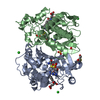
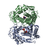
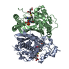
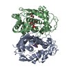
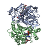
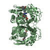
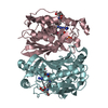
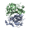
| Title | Crystal structure of White Spot Syndrome Virus Thymidylate Synthase - ternary complex with Methotrexate and dUMP | ||||||
 Components Components | Thymidylate Synthase | ||||||
 Keywords Keywords | TRANSFERASE / Thymidylate Synthase / BIOSYNTHETIC PROTEIN | ||||||
| Function / homology |  Function and homology informationthymidylate synthase / thymidylate synthase activity / dTMP biosynthetic process / dihydrofolate reductase activity / methylation / cytosol Function and homology informationthymidylate synthase / thymidylate synthase activity / dTMP biosynthetic process / dihydrofolate reductase activity / methylation / cytosolSimilarity search - Function | ||||||
| Biological species |  White spot syndrome virus White spot syndrome virus | ||||||
| Method | X-RAY DIFFRACTION / SYNCHROTRON / MOLECULAR REPLACEMENT / Resolution: 2.75 Å | ||||||
 Authors Authors | Panchal, N.V. / Kumar, S. / Shaikh, N. / Vasudevan, D. | ||||||
| Funding support |  India, 1items India, 1items
| ||||||
 Citation Citation | Journal: Int.J.Biol.Macromol. / Year: 2021 Title: Structure analysis of thymidylate synthase from white spot syndrome virus reveals WSSV-specific structural elements. Authors: Panchal, V. / Kumar, S. / Hossain, S.N. / Vasudevan, D. | ||||||
| History |
|
- Structure visualization
| Structure viewer | Molecule: MolmilJmol/JSmol |
|---|
- Downloads & links
-Download
| PDBx/mmCIF format | 7cca.cif.gz | 132.4 KB | Display | PDBx/mmCIF format |
|---|---|---|---|---|
| PDB format | pdb7cca.ent.gz | 101 KB | Display | PDB format |
| PDBx/mmJSON format | 7cca.json.gz | Tree view | PDBx/mmJSON format | |
| Others |  Other downloads Other downloads |
-Validation report
| Arichive directory | https://data.pdbj.org/pub/pdb/validation_reports/cc/7ccaftp://data.pdbj.org/pub/pdb/validation_reports/cc/7cca | HTTPS FTP |
|---|
-Related structure data
| Related structure data | 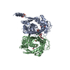 7cc8SC S: Starting model for refinement C: citing same article ( |
|---|---|
| Similar structure data |


-Links
 PDBj
PDBj
- Assembly
| Deposited unit | 
| ||||||||||||||||||||||||||||||
|---|---|---|---|---|---|---|---|---|---|---|---|---|---|---|---|---|---|---|---|---|---|---|---|---|---|---|---|---|---|---|---|
| 1 |
| ||||||||||||||||||||||||||||||
| Unit cell |
| ||||||||||||||||||||||||||||||
| Noncrystallographic symmetry (NCS) | NCS domain:
NCS domain segments: Component-ID: 1 / Ens-ID: 1 / Beg auth comp-ID: MET / Beg label comp-ID: MET / End auth comp-ID: LYS / End label comp-ID: LYS / Refine code: 1 / Auth seq-ID: 1 - 286 / Label seq-ID: 1 - 286
NCS oper:
|
-Components
| #1: Protein | / Wsv067 Mass: 34193.332 Da / Num. of mol.: 2 Source method: isolated from a genetically manipulated source Source: (gene. exp.) White spot syndrome virus (isolate Shrimp/China/Tongan/1996)Strain: isolate Shrimp/China/Tongan/1996 / Production host:  Escherichia coli BL21(DE3) (bacteria) / Strain (production host): BL21(DE3) / References: UniProt: Q77J90 Escherichia coli BL21(DE3) (bacteria) / Strain (production host): BL21(DE3) / References: UniProt: Q77J90#2: Chemical | Deoxyuridine monophosphate  Mass: 308.182 Da / Num. of mol.: 2 / Source method: obtained synthetically / Formula: C9H13N2O8P / Feature type: SUBJECT OF INVESTIGATION Mass: 308.182 Da / Num. of mol.: 2 / Source method: obtained synthetically / Formula: C9H13N2O8P / Feature type: SUBJECT OF INVESTIGATION#3: Chemical | Methotrexate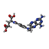  Mass: 454.439 Da / Num. of mol.: 2 / Source method: obtained synthetically / Formula: C20H22N8O5 / Feature type: SUBJECT OF INVESTIGATION / Comment: chemotherapy*YM Mass: 454.439 Da / Num. of mol.: 2 / Source method: obtained synthetically / Formula: C20H22N8O5 / Feature type: SUBJECT OF INVESTIGATION / Comment: chemotherapy*YM#4: Water | ChemComp-HOH / | Water Mass: 18.015 Da / Num. of mol.: 31 / Source method: isolated from a natural source / Formula: H2O Mass: 18.015 Da / Num. of mol.: 31 / Source method: isolated from a natural source / Formula: H2OHas ligand of interest | Y | |
|---|
-Experimental details
-Experiment
| Experiment | Method: X-RAY DIFFRACTION / Number of used crystals: 1 |
|---|
- Sample preparation
| Crystal | Density Matthews: 3.66 Å3/Da / Density % sol: 66.41 % |
|---|---|
| Crystal grow | Temperature: 291 K / Method: vapor diffusion, sitting drop / pH: 7 / Details: 1.1 M Ammonium tartrate dibasic |
-Data collection
| Diffraction | Mean temperature: 100 K / Serial crystal experiment: N |
|---|---|
| Diffraction source | Source: SYNCHROTRON / Site: ESRF  / Beamline: MASSIF-3 / Wavelength: 0.9677 Å / Beamline: MASSIF-3 / Wavelength: 0.9677 Å |
| Detector | Type: DECTRIS EIGER X 4M / Detector: PIXEL / Date: Mar 10, 2018 |
| Radiation | Protocol: SINGLE WAVELENGTH / Monochromatic (M) / Laue (L): M / Scattering type: x-ray |
| Radiation wavelength | Wavelength: 0.9677 Å / Relative weight: 1 |
| Reflection | Resolution: 2.75→45.36 Å / Num. obs: 24626 / % possible obs: 95.4 % / Redundancy: 4.6 % / CC1/2: 0.997 / Rmerge(I) obs: 0.087 / Net I/σ(I): 12.7 |
| Reflection shell | Resolution: 2.75→2.9 Å / Redundancy: 4.6 % / Rmerge(I) obs: 0.603 / Mean I/σ(I) obs: 2.4 / Num. unique obs: 3858 / CC1/2: 0.711 / % possible all: 98.3 |
- Processing
| Software |
| ||||||||||||||||||||||||||||||||||||||||||||||||||||||||||||
|---|---|---|---|---|---|---|---|---|---|---|---|---|---|---|---|---|---|---|---|---|---|---|---|---|---|---|---|---|---|---|---|---|---|---|---|---|---|---|---|---|---|---|---|---|---|---|---|---|---|---|---|---|---|---|---|---|---|---|---|---|---|
| Refinement | Method to determine structure: MOLECULAR REPLACEMENT Starting model: 7CC8 Resolution: 2.75→45.36 Å / Cor.coef. Fo:Fc: 0.95 / Cor.coef. Fo:Fc free: 0.929 / SU B: 10.61 / SU ML: 0.204 / Cross valid method: THROUGHOUT / σ(F): 0 / ESU R: 0.489 / ESU R Free: 0.286 / Stereochemistry target values: MAXIMUM LIKELIHOOD Details: HYDROGENS HAVE BEEN ADDED IN THE RIDING POSITIONS U VALUES : REFINED INDIVIDUALLY
| ||||||||||||||||||||||||||||||||||||||||||||||||||||||||||||
| Solvent computation | Ion probe radii: 0.8 Å / Shrinkage radii: 0.8 Å / VDW probe radii: 1.2 Å / Solvent model: MASK | ||||||||||||||||||||||||||||||||||||||||||||||||||||||||||||
| Displacement parameters | Biso max: 169.72 Å2 / Biso mean: 45.284 Å2 / Biso min: 21.83 Å2
| ||||||||||||||||||||||||||||||||||||||||||||||||||||||||||||
| Refinement step | Cycle: final / Resolution: 2.75→45.36 Å
| ||||||||||||||||||||||||||||||||||||||||||||||||||||||||||||
| Refine LS restraints |
| ||||||||||||||||||||||||||||||||||||||||||||||||||||||||||||
| Refine LS restraints NCS | Number: 2304 / Type: TIGHT THERMAL / Rms dev position: 6.68 Å / Weight position: 0.5 | ||||||||||||||||||||||||||||||||||||||||||||||||||||||||||||
| LS refinement shell | Resolution: 2.75→2.821 Å / Rfactor Rfree error: 0 / Total num. of bins used: 20
|
