 Movie
Movie Controller

[English] 日本語
 Yorodumi
- PDB-7c5f: Crystal Structure of Glyceraldehyde-3-phosphate dehydrogenase1 fr... -
+ Open data

- Basic information
| Entry | Database: PDB / ID: 7c5f | |||||||||
|---|---|---|---|---|---|---|---|---|---|---|
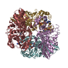
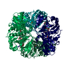
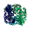
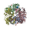
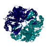
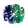
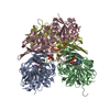
| Title | Crystal Structure of Glyceraldehyde-3-phosphate dehydrogenase1 from Escherichia coli at 1.88 Angstrom resolution | |||||||||
 Components Components | Glyceraldehyde-3-phosphate dehydrogenase Glyceraldehyde 3-phosphate dehydrogenase Glyceraldehyde 3-phosphate dehydrogenase | |||||||||
 Keywords Keywords | OXIDOREDUCTASE / EcGAPDH 1 / NAD | |||||||||
| Function / homology |  Function and homology informationOxidoreductases; Acting on the aldehyde or oxo group of donors; With NAD+ or NADP+ as acceptor / glyceraldehyde-3-phosphate dehydrogenase (NAD+) (phosphorylating) activity / glucose metabolic process / NAD binding / NADP binding / identical protein binding / cytoplasm Function and homology informationOxidoreductases; Acting on the aldehyde or oxo group of donors; With NAD+ or NADP+ as acceptor / glyceraldehyde-3-phosphate dehydrogenase (NAD+) (phosphorylating) activity / glucose metabolic process / NAD binding / NADP binding / identical protein binding / cytoplasmSimilarity search - Function | |||||||||
| Biological species |  Escherichia coli BL21 (bacteria) Escherichia coli BL21 (bacteria) | |||||||||
| Method | X-RAY DIFFRACTION / SYNCHROTRON / MOLECULAR REPLACEMENT / Resolution: 1.88 Å | |||||||||
 Authors Authors | Zhang, L. / Liu, M.R. / Yao, Y.C. / Bostrom, I.K. / Wang, Y.D. / Chen, A.Q. / Li, J.X. / Gu, S.H. / Ji, C.N. | |||||||||
| Funding support |  China, 2items China, 2items
| |||||||||
 Citation Citation | Journal: Acta Crystallogr.,Sect.F / Year: 2020 Title: Characterization and structure of glyceraldehyde-3-phosphate dehydrogenase type 1 from Escherichia coli. Authors: Zhang, L. / Liu, M.R. / Yao, Y.C. / Bostrom, I.K. / Wang, Y.D. / Chen, A.Q. / Li, J.X. / Gu, S.H. / Ji, C.N. | |||||||||
| History |
|
- Structure visualization
| Structure viewer | Molecule: MolmilJmol/JSmol |
|---|
- Downloads & links
-Download
| PDBx/mmCIF format | 7c5f.cif.gz | 273.4 KB | Display | PDBx/mmCIF format |
|---|---|---|---|---|
| PDB format | pdb7c5f.ent.gz | 219.7 KB | Display | PDB format |
| PDBx/mmJSON format | 7c5f.json.gz | Tree view | PDBx/mmJSON format | |
| Others |  Other downloads Other downloads |
-Validation report
| Arichive directory | https://data.pdbj.org/pub/pdb/validation_reports/c5/7c5fftp://data.pdbj.org/pub/pdb/validation_reports/c5/7c5f | HTTPS FTP |
|---|
-Related structure data
| Related structure data | 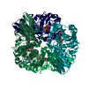 3k73S S: Starting model for refinement |
|---|---|
| Similar structure data |


-Links
 PDBj
PDBj
- Assembly
| Deposited unit | 
| ||||||||
|---|---|---|---|---|---|---|---|---|---|
| 1 |
| ||||||||
| Unit cell |
|
-Components
| #1: Protein | Glyceraldehyde 3-phosphate dehydrogenase Mass: 37887.922 Da / Num. of mol.: 4 Source method: isolated from a genetically manipulated source Source: (gene. exp.) Escherichia coli BL21(DE3) (bacteria) / Gene: ECBD_2224 / Production host: Escherichia coli BL21(DE3) (bacteria)References: UniProt: A0A140NCK4, Oxidoreductases; Acting on the aldehyde or oxo group of donors; With NAD+ or NADP+ as acceptor#2: Chemical | ChemComp-NAD / Nicotinamide adenine dinucleotide  Mass: 663.425 Da / Num. of mol.: 4 / Source method: isolated from a natural source / Formula: C21H27N7O14P2 / Feature type: SUBJECT OF INVESTIGATION / Comment: NAD*YM Mass: 663.425 Da / Num. of mol.: 4 / Source method: isolated from a natural source / Formula: C21H27N7O14P2 / Feature type: SUBJECT OF INVESTIGATION / Comment: NAD*YM#3: Chemical | ChemComp-PO4 / | Phosphate  Mass: 94.971 Da / Num. of mol.: 1 / Source method: obtained synthetically / Formula: PO4 / Feature type: SUBJECT OF INVESTIGATION Mass: 94.971 Da / Num. of mol.: 1 / Source method: obtained synthetically / Formula: PO4 / Feature type: SUBJECT OF INVESTIGATION#4: Water | ChemComp-HOH / | Water Mass: 18.015 Da / Num. of mol.: 568 / Source method: isolated from a natural source / Formula: H2O Mass: 18.015 Da / Num. of mol.: 568 / Source method: isolated from a natural source / Formula: H2OHas ligand of interest | Y | |
|---|
-Experimental details
-Experiment
| Experiment | Method: X-RAY DIFFRACTION / Number of used crystals: 1 |
|---|
- Sample preparation
| Crystal | Density Matthews: 2.26 Å3/Da / Density % sol: 45.59 % |
|---|---|
| Crystal grow | Temperature: 293 K / Method: vapor diffusion, hanging drop / pH: 4.6 Details: 100mM sodium acetate pH4.6, 30%(w/v) PEG 400, 200mM Ca(OAC)2 |
-Data collection
| Diffraction | Mean temperature: 100 K / Serial crystal experiment: N |
|---|---|
| Diffraction source | Source: SYNCHROTRON / Site: SSRF / Beamline: BL17U1 / Wavelength: 0.97915 Å |
| Detector | Type: ADSC QUANTUM 315 / Detector: CCD / Date: Jan 6, 2016 |
| Radiation | Protocol: SINGLE WAVELENGTH / Monochromatic (M) / Laue (L): M / Scattering type: x-ray |
| Radiation wavelength | Wavelength: 0.97915 Å / Relative weight: 1 |
| Reflection | Resolution: 1.88→50 Å / Num. obs: 114194 / % possible obs: 99.9 % / Redundancy: 8.9 % / Rmerge(I) obs: 0.2 / Net I/σ(I): 2 |
| Reflection shell | Resolution: 1.88→1.91 Å / Redundancy: 9.8 % / Rmerge(I) obs: 1.8 / Mean I/σ(I) obs: 13.5 / Num. unique obs: 114194 / CC1/2: 0.526 / Rpim(I) all: 0.637 / % possible all: 100 |
- Processing
| Software |
| ||||||||||||||||
|---|---|---|---|---|---|---|---|---|---|---|---|---|---|---|---|---|---|
| Refinement | Method to determine structure: MOLECULAR REPLACEMENT Starting model: 3K73 Resolution: 1.88→37.81 Å / Cross valid method: FREE R-VALUE
| ||||||||||||||||
| Refinement step | Cycle: LAST / Resolution: 1.88→37.81 Å
| ||||||||||||||||
| LS refinement shell | Highest resolution: 1.88 Å /
|
