 Movie
Movie Controller

[English] 日本語
 Yorodumi
- PDB-7bqw: Crystal structure of Methionine gamma-lyase from Fusobacterium nu... -
+ Open data

- Basic information
| Entry | Database: PDB / ID: 7bqw | |||||||||
|---|---|---|---|---|---|---|---|---|---|---|
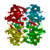
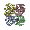
| Title | Crystal structure of Methionine gamma-lyase from Fusobacterium nucleatum | |||||||||
 Components Components | L-methionine gamma-lyase | |||||||||
 Keywords Keywords |  LYASE / Methionine gamma-lyase / Fn1419 / Fusobacterium nucleatum LYASE / Methionine gamma-lyase / Fn1419 / Fusobacterium nucleatum | |||||||||
| Function / homology |  Function and homology information Function and homology informationL-cysteine desulfidase / homocysteine desulfhydrase / homocysteine desulfhydrase activity / carbon-sulfur lyase activity / methionine gamma-lyase / methionine gamma-lyase activity / L-cysteine desulfhydrase activity / transsulfuration / pyridoxal phosphate binding / cytoplasmSimilarity search - Function | |||||||||
| Biological species |  Fusobacterium nucleatum subsp. nucleatum ATCC 25586 (bacteria) Fusobacterium nucleatum subsp. nucleatum ATCC 25586 (bacteria) | |||||||||
| Method | X-RAY DIFFRACTION / SYNCHROTRON / MOLECULAR REPLACEMENT / Resolution: 2.5 Å | |||||||||
 Authors Authors | Lan, J. / Chen, Y. / Liu, W. / Xu, Y. | |||||||||
| Funding support |  China, 2items China, 2items
| |||||||||
 Citation Citation | Journal: To Be Published Title: Crystal structure of Methionine gamma-lyase from Fusobacterium nucleatum Authors: Lan, J. / Liu, W. / Chen, Y. / Xu, Y. | |||||||||
| History |
|
- Structure visualization
| Structure viewer | Molecule: MolmilJmol/JSmol |
|---|
- Downloads & links
-Download
| PDBx/mmCIF format | 7bqw.cif.gz | 340.2 KB | Display | PDBx/mmCIF format |
|---|---|---|---|---|
| PDB format | pdb7bqw.ent.gz | 221.7 KB | Display | PDB format |
| PDBx/mmJSON format | 7bqw.json.gz | Tree view | PDBx/mmJSON format | |
| Others |  Other downloads Other downloads |
-Validation report
| Arichive directory | https://data.pdbj.org/pub/pdb/validation_reports/bq/7bqwftp://data.pdbj.org/pub/pdb/validation_reports/bq/7bqw | HTTPS FTP |
|---|
-Related structure data
| Related structure data |  5dx5S S: Starting model for refinement |
|---|---|
| Similar structure data |


-Links
 PDBj
PDBj- Assembly
| Deposited unit | 
| ||||||||||||
|---|---|---|---|---|---|---|---|---|---|---|---|---|---|
| 1 |
| ||||||||||||
| Unit cell |
|
-Components
| #1: Protein | Mass: 43357.762 Da / Num. of mol.: 4 Source method: isolated from a genetically manipulated source Source: (gene. exp.) Fusobacterium nucleatum subsp. nucleatum ATCC 25586 (bacteria)Gene: FN1419 / Production host: Escherichia coli (E. coli)References: UniProt: Q8RDT4, methionine gamma-lyase, homocysteine desulfhydrase, L-cysteine desulfidase#2: Water | ChemComp-HOH / | Water Mass: 18.015 Da / Num. of mol.: 135 / Source method: isolated from a natural source / Formula: H2O Mass: 18.015 Da / Num. of mol.: 135 / Source method: isolated from a natural source / Formula: H2O |
|---|
-Experimental details
-Experiment
| Experiment | Method: X-RAY DIFFRACTION / Number of used crystals: 1 |
|---|
- Sample preparation
| Crystal | Density Matthews: 2.28 Å3/Da / Density % sol: 46.05 % |
|---|---|
| Crystal grow | Temperature: 295 K / Method: vapor diffusion, sitting drop Details: 20% PEG 5000, 0.2M sodium formate, 0.1M BICINE pH 8.5 |
-Data collection
| Diffraction | Mean temperature: 100 K / Serial crystal experiment: N |
|---|---|
| Diffraction source | Source: SYNCHROTRON / Site: SSRF / Beamline: BL19U1 / Wavelength: 0.9789 Å |
| Detector | Type: ADSC QUANTUM 315r / Detector: CCD / Date: Jan 9, 2020 |
| Radiation | Protocol: SINGLE WAVELENGTH / Monochromatic (M) / Laue (L): M / Scattering type: x-ray |
| Radiation wavelength | Wavelength: 0.9789 Å / Relative weight: 1 |
| Reflection | Resolution: 2.5→50 Å / Num. obs: 56011 / % possible obs: 99.8 % / Redundancy: 3.9 % / Biso Wilson estimate: 47.74 Å2 / Rsym value: 0.07 / Net I/σ(I): 23.77 |
| Reflection shell | Resolution: 2.5→2.59 Å / Num. unique obs: 55899 / Rsym value: 0.32 |
- Processing
| Software |
| |||||||||||||||||||||||||||||||||||||||||||||||||||||||||||||||||||||||||||||||||||||||||||||||||||||||||||||||||||||||||||||||||||||||||||||||||||
|---|---|---|---|---|---|---|---|---|---|---|---|---|---|---|---|---|---|---|---|---|---|---|---|---|---|---|---|---|---|---|---|---|---|---|---|---|---|---|---|---|---|---|---|---|---|---|---|---|---|---|---|---|---|---|---|---|---|---|---|---|---|---|---|---|---|---|---|---|---|---|---|---|---|---|---|---|---|---|---|---|---|---|---|---|---|---|---|---|---|---|---|---|---|---|---|---|---|---|---|---|---|---|---|---|---|---|---|---|---|---|---|---|---|---|---|---|---|---|---|---|---|---|---|---|---|---|---|---|---|---|---|---|---|---|---|---|---|---|---|---|---|---|---|---|---|---|---|---|
| Refinement | Method to determine structure: MOLECULAR REPLACEMENT Starting model: 5DX5 Resolution: 2.5→48.75 Å / SU ML: 0.3658 / Cross valid method: FREE R-VALUE / σ(F): 1.34 / Phase error: 29.5913
| |||||||||||||||||||||||||||||||||||||||||||||||||||||||||||||||||||||||||||||||||||||||||||||||||||||||||||||||||||||||||||||||||||||||||||||||||||
| Solvent computation | Shrinkage radii: 0.9 Å / VDW probe radii: 1.11 Å | |||||||||||||||||||||||||||||||||||||||||||||||||||||||||||||||||||||||||||||||||||||||||||||||||||||||||||||||||||||||||||||||||||||||||||||||||||
| Displacement parameters | Biso mean: 52.66 Å2 | |||||||||||||||||||||||||||||||||||||||||||||||||||||||||||||||||||||||||||||||||||||||||||||||||||||||||||||||||||||||||||||||||||||||||||||||||||
| Refinement step | Cycle: LAST / Resolution: 2.5→48.75 Å
| |||||||||||||||||||||||||||||||||||||||||||||||||||||||||||||||||||||||||||||||||||||||||||||||||||||||||||||||||||||||||||||||||||||||||||||||||||
| Refine LS restraints |
| |||||||||||||||||||||||||||||||||||||||||||||||||||||||||||||||||||||||||||||||||||||||||||||||||||||||||||||||||||||||||||||||||||||||||||||||||||
| LS refinement shell |
|
