 Movie
Movie Controller

[English] 日本語
 Yorodumi
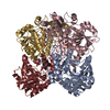
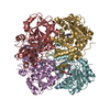
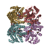
- PDB-6khq: bacterial cystathionine gamma-lyase MccB of Staphylococcus aureus... -
+ Open data

- Basic information
| Entry | Database: PDB / ID: 6khq | ||||||
|---|---|---|---|---|---|---|---|
| Title | bacterial cystathionine gamma-lyase MccB of Staphylococcus aureus with cofactor PLP | ||||||
 Components Components | Cystathionine gamma lyase | ||||||
 Keywords Keywords |  LYASE / Cystathionine gamma lyase / CGL / MccB / yhrB / PLP / Pyridoxal Phosphate / Cysteine synthesis LYASE / Cystathionine gamma lyase / CGL / MccB / yhrB / PLP / Pyridoxal Phosphate / Cysteine synthesis | ||||||
| Function / homology |  Function and homology information Function and homology information | ||||||
| Biological species |  Staphylococcus aureus subsp. aureus Mu50 (bacteria) Staphylococcus aureus subsp. aureus Mu50 (bacteria) | ||||||
| Method | X-RAY DIFFRACTION / SYNCHROTRON / MOLECULAR REPLACEMENT / Resolution: 2.3 Å | ||||||
 Authors Authors | Ha, N.-C. / Lee, D. | ||||||
 Citation Citation | Journal: Crystals / Year: 2019 Title: Crystal Structure of Bacterial Cystathionine Gamma-Lyase in The Cysteine Biosynthesis Pathway of Staphylococcus aureus Authors: Lee, D. / Jeong, S. / Ahn, J. / Ha, N.-C. / Kwon, A.-R. | ||||||
| History |
|
- Structure visualization
| Structure viewer | Molecule: MolmilJmol/JSmol |
|---|
- Downloads & links
-Download
| PDBx/mmCIF format | 6khq.cif.gz | 191 KB | Display | PDBx/mmCIF format |
|---|---|---|---|---|
| PDB format | pdb6khq.ent.gz | 121 KB | Display | PDB format |
| PDBx/mmJSON format | 6khq.json.gz | Tree view | PDBx/mmJSON format | |
| Others |  Other downloads Other downloads |
-Validation report
| Arichive directory | https://data.pdbj.org/pub/pdb/validation_reports/kh/6khqftp://data.pdbj.org/pub/pdb/validation_reports/kh/6khq | HTTPS FTP |
|---|
-Related structure data
| Related structure data |  6kgzSC S: Starting model for refinement C: citing same article ( |
|---|---|
| Similar structure data |


-Links
 PDBj
PDBj- Assembly
| Deposited unit | 
| ||||||||||||
|---|---|---|---|---|---|---|---|---|---|---|---|---|---|
| 1 | 
| ||||||||||||
| Unit cell |
| ||||||||||||
| Components on special symmetry positions |
|
-Components
| #1: Protein | Mass: 43565.016 Da / Num. of mol.: 2 Source method: isolated from a genetically manipulated source Source: (gene. exp.) Staphylococcus aureus subsp. aureus Mu50 (bacteria)Strain: Mu50 / Gene: yrhB, SAV0460 / Production host: Escherichia coli BL21(DE3) (bacteria) / Strain (production host): BL21(DE3) / References: UniProt: A0A0H3JQ19, cystathionine gamma-lyase#2: Water | ChemComp-HOH / | Water Mass: 18.015 Da / Num. of mol.: 139 / Source method: isolated from a natural source / Formula: H2O Mass: 18.015 Da / Num. of mol.: 139 / Source method: isolated from a natural source / Formula: H2OHas ligand of interest | Y | |
|---|
-Experimental details
-Experiment
| Experiment | Method: X-RAY DIFFRACTION / Number of used crystals: 1 |
|---|
- Sample preparation
| Crystal | Density Matthews: 2.65 Å3/Da / Density % sol: 53.55 % |
|---|---|
| Crystal grow | Temperature: 288 K / Method: vapor diffusion, hanging drop / pH: 4 / Details: PEG 400, Sodium Acetate |
-Data collection
| Diffraction | Mean temperature: 100 K / Serial crystal experiment: N |
|---|---|
| Diffraction source | Source: SYNCHROTRON / Site: PAL/PLS  / Beamline: 5C (4A) / Wavelength: 1.00003 Å / Beamline: 5C (4A) / Wavelength: 1.00003 Å |
| Detector | Type: DECTRIS EIGER X 9M / Detector: PIXEL / Date: Jun 19, 2019 |
| Radiation | Protocol: SINGLE WAVELENGTH / Monochromatic (M) / Laue (L): M / Scattering type: x-ray |
| Radiation wavelength | Wavelength: 1.00003 Å / Relative weight: 1 |
| Reflection | Resolution: 2.3→50 Å / Num. obs: 39074 / % possible obs: 99.5 % / Redundancy: 14.1 % / Biso Wilson estimate: 26.43 Å2 / CC1/2: 1 / Rpim(I) all: 0.017 / Rrim(I) all: 0.072 / Net I/σ(I): 28.11 |
| Reflection shell | Resolution: 2.3→2.34 Å / Redundancy: 7.5 % / Mean I/σ(I) obs: 3.92 / Num. unique obs: 1914 / CC1/2: 0.701 / Rpim(I) all: 0.126 / Rrim(I) all: 0.373 / % possible all: 98.7 |
- Processing
| Software |
| |||||||||||||||||||||||||||||||||||||||||||||||||||||||||||||||||||||||||||||||||||||||||||||||||||||||||
|---|---|---|---|---|---|---|---|---|---|---|---|---|---|---|---|---|---|---|---|---|---|---|---|---|---|---|---|---|---|---|---|---|---|---|---|---|---|---|---|---|---|---|---|---|---|---|---|---|---|---|---|---|---|---|---|---|---|---|---|---|---|---|---|---|---|---|---|---|---|---|---|---|---|---|---|---|---|---|---|---|---|---|---|---|---|---|---|---|---|---|---|---|---|---|---|---|---|---|---|---|---|---|---|---|---|---|
| Refinement | Method to determine structure: MOLECULAR REPLACEMENT Starting model: 6KGZ Resolution: 2.3→42.2 Å / SU ML: 0.2616 / Cross valid method: FREE R-VALUE / σ(F): 1.53 / Phase error: 21.8161
| |||||||||||||||||||||||||||||||||||||||||||||||||||||||||||||||||||||||||||||||||||||||||||||||||||||||||
| Solvent computation | Shrinkage radii: 0.9 Å / VDW probe radii: 1.11 Å | |||||||||||||||||||||||||||||||||||||||||||||||||||||||||||||||||||||||||||||||||||||||||||||||||||||||||
| Displacement parameters | Biso mean: 30.28 Å2 | |||||||||||||||||||||||||||||||||||||||||||||||||||||||||||||||||||||||||||||||||||||||||||||||||||||||||
| Refinement step | Cycle: LAST / Resolution: 2.3→42.2 Å
| |||||||||||||||||||||||||||||||||||||||||||||||||||||||||||||||||||||||||||||||||||||||||||||||||||||||||
| Refine LS restraints |
| |||||||||||||||||||||||||||||||||||||||||||||||||||||||||||||||||||||||||||||||||||||||||||||||||||||||||
| LS refinement shell |
|
