 Movie
Movie Controller

[English] 日本語
 Yorodumi
- PDB-7b4i: Thermostable omega transaminase PjTA-R6 variant W58G engineered f... -
+ Open data

- Basic information
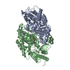
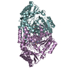
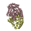
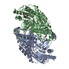
| Entry | Database: PDB / ID: 7b4i | |||||||||
|---|---|---|---|---|---|---|---|---|---|---|
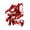
| Title | Thermostable omega transaminase PjTA-R6 variant W58G engineered for asymmetric synthesis of enantiopure bulky amines | |||||||||
 Components Components | Aspartate aminotransferase family protein | |||||||||
 Keywords Keywords |  TRANSFERASE / Aminotransferase / Transaminase / Amine synthesis / Enantioselective / Thermostable / Engineered TRANSFERASE / Aminotransferase / Transaminase / Amine synthesis / Enantioselective / Thermostable / Engineered | |||||||||
| Function / homology |  Function and homology information Function and homology information | |||||||||
| Biological species |  Pseudomonas sp. (bacteria) Pseudomonas sp. (bacteria) | |||||||||
| Method | X-RAY DIFFRACTION / SYNCHROTRON / FOURIER SYNTHESIS / Resolution: 1.7 Å | |||||||||
 Authors Authors | Capra, N. / Rozeboom, H.J. / Thunnissen, A.M.W.H. / Janssen, D.B. | |||||||||
| Funding support |  Netherlands, 1items Netherlands, 1items
| |||||||||
 Citation Citation | Journal: Acs Catalysis / Year: 2021 Title: Computational Redesign of an omega-Transaminase from Pseudomonas jessenii for Asymmetric Synthesis of Enantiopure Bulky Amines. Authors: Meng, Q. / Ramirez-Palacios, C. / Capra, N. / Hooghwinkel, M.E. / Thallmair, S. / Rozeboom, H.J. / Thunnissen, A.W.H. / Wijma, H.J. / Marrink, S.J. / Janssen, D.B. | |||||||||
| History |
|
- Structure visualization
| Structure viewer | Molecule: MolmilJmol/JSmol |
|---|
- Downloads & links
-Download
| PDBx/mmCIF format | 7b4i.cif.gz | 351.5 KB | Display | PDBx/mmCIF format |
|---|---|---|---|---|
| PDB format | pdb7b4i.ent.gz | Display | PDB format | |
| PDBx/mmJSON format | 7b4i.json.gz | Tree view | PDBx/mmJSON format | |
| Others |  Other downloads Other downloads |
-Validation report
| Arichive directory | https://data.pdbj.org/pub/pdb/validation_reports/b4/7b4iftp://data.pdbj.org/pub/pdb/validation_reports/b4/7b4i | HTTPS FTP |
|---|
-Related structure data


-Links
 PDBj
PDBj- Assembly
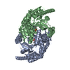
| Deposited unit | 
| ||||||||
|---|---|---|---|---|---|---|---|---|---|
| 1 |
| ||||||||
| Unit cell |
| ||||||||
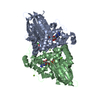
| Noncrystallographic symmetry (NCS) | NCS domain: (Details: Chains AAA BBB) |
-Components
| #1: Protein | Mass: 50628.684 Da / Num. of mol.: 2 / Mutation: W58G Source method: isolated from a genetically manipulated source Source: (gene. exp.) Pseudomonas sp. (bacteria) / Gene: CMK94_18730, DIU04_17820 / Production host: Escherichia coli BL21(DE3) (bacteria) / References: UniProt: A0A2D8IND4#2: Chemical | Pyridoxal phosphate  Mass: 247.142 Da / Num. of mol.: 2 / Source method: obtained synthetically / Formula: C8H10NO6P / Feature type: SUBJECT OF INVESTIGATION Mass: 247.142 Da / Num. of mol.: 2 / Source method: obtained synthetically / Formula: C8H10NO6P / Feature type: SUBJECT OF INVESTIGATION#3: Chemical | ChemComp-SIN / | Succinic acid  Mass: 118.088 Da / Num. of mol.: 1 / Source method: obtained synthetically / Formula: C4H6O4 Mass: 118.088 Da / Num. of mol.: 1 / Source method: obtained synthetically / Formula: C4H6O4#4: Water | ChemComp-HOH / | Water Mass: 18.015 Da / Num. of mol.: 428 / Source method: isolated from a natural source / Formula: H2O Mass: 18.015 Da / Num. of mol.: 428 / Source method: isolated from a natural source / Formula: H2OHas ligand of interest | N | |
|---|
-Experimental details
-Experiment
| Experiment | Method: X-RAY DIFFRACTION / Number of used crystals: 1 |
|---|
- Sample preparation
| Crystal | Density Matthews: 2.83 Å3/Da / Density % sol: 56.54 % |
|---|---|
| Crystal grow | Temperature: 293 K / Method: vapor diffusion, hanging drop / pH: 7.6 Details: Drops were prepared by mixing 1 ul of protein solution (~10 mg/ml in 20 mM HEPES pH 7.5, 100 mM NaCl, 20 uM PLP buffer) with 1 ul of reservoir solution. Reservoir contained 0.7-1M succinic ...Details: Drops were prepared by mixing 1 ul of protein solution (~10 mg/ml in 20 mM HEPES pH 7.5, 100 mM NaCl, 20 uM PLP buffer) with 1 ul of reservoir solution. Reservoir contained 0.7-1M succinic acid pH 7.6. Crystals appeared after 48h. |
-Data collection
| Diffraction | Mean temperature: 100 K / Serial crystal experiment: N | ||||||||||||||||||
|---|---|---|---|---|---|---|---|---|---|---|---|---|---|---|---|---|---|---|---|
| Diffraction source | Source: SYNCHROTRON / Site: DIAMOND  / Beamline: I24 / Wavelength: 0.9686 Å / Beamline: I24 / Wavelength: 0.9686 Å | ||||||||||||||||||
| Detector | Type: DECTRIS PILATUS 6M / Detector: PIXEL / Date: Mar 4, 2020 | ||||||||||||||||||
| Radiation | Protocol: SINGLE WAVELENGTH / Monochromatic (M) / Laue (L): M / Scattering type: x-ray | ||||||||||||||||||
| Radiation wavelength | Wavelength: 0.9686 Å / Relative weight: 1 | ||||||||||||||||||
| Reflection | Resolution: 1.7→98 Å / Num. obs: 123358 / % possible obs: 99.9 % / Redundancy: 5.5 % / CC1/2: 0.999 / Rmerge(I) obs: 0.038 / Rpim(I) all: 0.027 / Rrim(I) all: 0.046 / Net I/σ(I): 17.4 | ||||||||||||||||||
| Reflection shell | Diffraction-ID: 1 / Redundancy: 5.5 %
|
- Processing
| Software |
| |||||||||||||||||||||||||||||||||||||||||||||||||||||||||||||||||||||||||||||||||||||||||||||||||||||||||||||||||||||||||||||||||||||||||||||||||||||||||||
|---|---|---|---|---|---|---|---|---|---|---|---|---|---|---|---|---|---|---|---|---|---|---|---|---|---|---|---|---|---|---|---|---|---|---|---|---|---|---|---|---|---|---|---|---|---|---|---|---|---|---|---|---|---|---|---|---|---|---|---|---|---|---|---|---|---|---|---|---|---|---|---|---|---|---|---|---|---|---|---|---|---|---|---|---|---|---|---|---|---|---|---|---|---|---|---|---|---|---|---|---|---|---|---|---|---|---|---|---|---|---|---|---|---|---|---|---|---|---|---|---|---|---|---|---|---|---|---|---|---|---|---|---|---|---|---|---|---|---|---|---|---|---|---|---|---|---|---|---|---|---|---|---|---|---|---|---|
| Refinement | Method to determine structure: FOURIER SYNTHESIS / Resolution: 1.7→98 Å / Cor.coef. Fo:Fc: 0.974 / Cor.coef. Fo:Fc free: 0.968 / SU B: 2.005 / SU ML: 0.064 / Cross valid method: FREE R-VALUE / ESU R: 0.087 / ESU R Free: 0.083 Details: Hydrogens have been added in their riding positions
| |||||||||||||||||||||||||||||||||||||||||||||||||||||||||||||||||||||||||||||||||||||||||||||||||||||||||||||||||||||||||||||||||||||||||||||||||||||||||||
| Solvent computation | Ion probe radii: 0.8 Å / Shrinkage radii: 0.8 Å / VDW probe radii: 1.2 Å / Solvent model: MASK BULK SOLVENT | |||||||||||||||||||||||||||||||||||||||||||||||||||||||||||||||||||||||||||||||||||||||||||||||||||||||||||||||||||||||||||||||||||||||||||||||||||||||||||
| Displacement parameters | Biso mean: 30.6 Å2
| |||||||||||||||||||||||||||||||||||||||||||||||||||||||||||||||||||||||||||||||||||||||||||||||||||||||||||||||||||||||||||||||||||||||||||||||||||||||||||
| Refinement step | Cycle: LAST / Resolution: 1.7→98 Å
| |||||||||||||||||||||||||||||||||||||||||||||||||||||||||||||||||||||||||||||||||||||||||||||||||||||||||||||||||||||||||||||||||||||||||||||||||||||||||||
| Refine LS restraints |
| |||||||||||||||||||||||||||||||||||||||||||||||||||||||||||||||||||||||||||||||||||||||||||||||||||||||||||||||||||||||||||||||||||||||||||||||||||||||||||
| LS refinement shell |
|
