 Movie
Movie Controller

[English] 日本語
 Yorodumi
- PDB-7b1w: Crystal structure of plastidial ribulose epimerase RPE1 from the ... -
+ Open data

- Basic information
| Entry | Database: PDB / ID: 7b1w | ||||||||||||
|---|---|---|---|---|---|---|---|---|---|---|---|---|---|
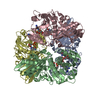
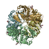
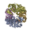
| Title | Crystal structure of plastidial ribulose epimerase RPE1 from the model alga Chlamydomonas reinhardtii | ||||||||||||
 Components Components | Ribulose-phosphate 3-epimerase Phosphopentose epimerase Phosphopentose epimerase | ||||||||||||
 Keywords Keywords | ISOMERASE / Calvin-Benson cycle / photosynthesis / ribulose / ribose / epimerase / chloroplast / carbon fixation | ||||||||||||
| Function / homology |  Function and homology information: / ribulose-phosphate 3-epimerase / D-ribulose-phosphate 3-epimerase activity / stromule / pentose catabolic process / pentose-phosphate shunt, non-oxidative branch / response to nematode / thylakoid / apoplast / chloroplast envelope ...: / ribulose-phosphate 3-epimerase / D-ribulose-phosphate 3-epimerase activity / stromule / pentose catabolic process / pentose-phosphate shunt, non-oxidative branch / response to nematode / thylakoid / apoplast / chloroplast envelope / chloroplast stroma / response to cold / metal ion binding / cytosol Function and homology information: / ribulose-phosphate 3-epimerase / D-ribulose-phosphate 3-epimerase activity / stromule / pentose catabolic process / pentose-phosphate shunt, non-oxidative branch / response to nematode / thylakoid / apoplast / chloroplast envelope ...: / ribulose-phosphate 3-epimerase / D-ribulose-phosphate 3-epimerase activity / stromule / pentose catabolic process / pentose-phosphate shunt, non-oxidative branch / response to nematode / thylakoid / apoplast / chloroplast envelope / chloroplast stroma / response to cold / metal ion binding / cytosolSimilarity search - Function | ||||||||||||
| Biological species |   Chlamydomonas reinhardtii (plant) Chlamydomonas reinhardtii (plant) | ||||||||||||
| Method | X-RAY DIFFRACTION / SYNCHROTRON / MOLECULAR REPLACEMENT / Resolution: 1.935 Å | ||||||||||||
 Authors Authors | Henri, J. / Zaffagnini, M. / Tedesco, D. / Crozet, P. / Lemaire, S.D. | ||||||||||||
| Funding support |  France, 3items France, 3items
| ||||||||||||
 Citation Citation | Journal: Plant Physiol. / Year: 2023 Title: Characterization of chloroplast ribulose-5-phosphate-3-epimerase from the microalga Chlamydomonas reinhardtii. Authors: Meloni, M. / Fanti, S. / Tedesco, D. / Gurrieri, L. / Trost, P. / Fermani, S. / Lemaire, S.D. / Zaffagnini, M. / Henri, J. | ||||||||||||
| History |
|
- Structure visualization
| Structure viewer | Molecule: MolmilJmol/JSmol |
|---|
- Downloads & links
-Download
| PDBx/mmCIF format | 7b1w.cif.gz | 552.3 KB | Display | PDBx/mmCIF format |
|---|---|---|---|---|
| PDB format | pdb7b1w.ent.gz | 450.8 KB | Display | PDB format |
| PDBx/mmJSON format | 7b1w.json.gz | Tree view | PDBx/mmJSON format | |
| Others |  Other downloads Other downloads |
-Validation report
| Arichive directory | https://data.pdbj.org/pub/pdb/validation_reports/b1/7b1wftp://data.pdbj.org/pub/pdb/validation_reports/b1/7b1w | HTTPS FTP |
|---|
-Related structure data


-Links
 PDBj
PDBj
- Assembly
| Deposited unit | 
| ||||||||
|---|---|---|---|---|---|---|---|---|---|
| 1 | 
| ||||||||
| 2 | 
| ||||||||
| Unit cell |
|
-Components
| #1: Protein | Phosphopentose epimerase Mass: 26811.033 Da / Num. of mol.: 12 Source method: isolated from a genetically manipulated source Source: (gene. exp.) Chlamydomonas reinhardtii (plant) / Gene: RPE1, CHLRE_12g511900v5, CHLREDRAFT_135614 / Production host:  Escherichia coli BL21(DE3) (bacteria) / References: UniProt: A8IKW6, ribulose-phosphate 3-epimerase Escherichia coli BL21(DE3) (bacteria) / References: UniProt: A8IKW6, ribulose-phosphate 3-epimerase#2: Chemical | ChemComp-ZN /   Mass: 65.409 Da / Num. of mol.: 12 / Source method: isolated from a natural source / Formula: Zn Mass: 65.409 Da / Num. of mol.: 12 / Source method: isolated from a natural source / Formula: Zn#3: Water | ChemComp-HOH / | Water Mass: 18.015 Da / Num. of mol.: 1739 / Source method: isolated from a natural source / Formula: H2O Mass: 18.015 Da / Num. of mol.: 1739 / Source method: isolated from a natural source / Formula: H2OHas ligand of interest | N | |
|---|
-Experimental details
-Experiment
| Experiment | Method: X-RAY DIFFRACTION / Number of used crystals: 1 |
|---|
- Sample preparation
| Crystal | Density Matthews: 2.4 Å3/Da / Density % sol: 48.8 % |
|---|---|
| Crystal grow | Temperature: 293 K / Method: vapor diffusion, sitting drop Details: Calcium acetate 160 mmol/L Sodium cacodylate pH 6.5 80 mmol/L Glycerol 20 % Polyethylene glycol 8000 14.4 % |
-Data collection
| Diffraction | Mean temperature: 100 K / Serial crystal experiment: N |
|---|---|
| Diffraction source | Source: SYNCHROTRON / Site: ESRF / Beamline: MASSIF-3 / Wavelength: 0.9677 Å |
| Detector | Type: DECTRIS EIGER X 4M / Detector: PIXEL / Date: Mar 17, 2017 |
| Radiation | Protocol: SINGLE WAVELENGTH / Monochromatic (M) / Laue (L): M / Scattering type: x-ray |
| Radiation wavelength | Wavelength: 0.9677 Å / Relative weight: 1 |
| Reflection | Resolution: 1.935→49.29 Å / Num. obs: 229414 / % possible obs: 98.65 % / Redundancy: 7.2 % / Biso Wilson estimate: 30.3 Å2 / CC1/2: 0.997 / Net I/σ(I): 9.79 |
| Reflection shell | Resolution: 1.935→2.005 Å / Num. unique obs: 21621 / CC1/2: 0.616 |
- Processing
| Software |
| ||||||||||||||||||||||||||||||||||||||||||||||||||||||||||||||||||||||||||||||||||||||||||||||||||||||||||||
|---|---|---|---|---|---|---|---|---|---|---|---|---|---|---|---|---|---|---|---|---|---|---|---|---|---|---|---|---|---|---|---|---|---|---|---|---|---|---|---|---|---|---|---|---|---|---|---|---|---|---|---|---|---|---|---|---|---|---|---|---|---|---|---|---|---|---|---|---|---|---|---|---|---|---|---|---|---|---|---|---|---|---|---|---|---|---|---|---|---|---|---|---|---|---|---|---|---|---|---|---|---|---|---|---|---|---|---|---|---|
| Refinement | Method to determine structure: MOLECULAR REPLACEMENT Starting model: Homology model Resolution: 1.935→49.289 Å / SU ML: 0.25 / Cross valid method: THROUGHOUT / σ(F): 1.33 / Phase error: 23.56 / Stereochemistry target values: ML
| ||||||||||||||||||||||||||||||||||||||||||||||||||||||||||||||||||||||||||||||||||||||||||||||||||||||||||||
| Solvent computation | Shrinkage radii: 0.9 Å / VDW probe radii: 1.11 Å / Solvent model: FLAT BULK SOLVENT MODEL | ||||||||||||||||||||||||||||||||||||||||||||||||||||||||||||||||||||||||||||||||||||||||||||||||||||||||||||
| Displacement parameters | Biso max: 83.91 Å2 / Biso mean: 34.7765 Å2 / Biso min: 15.65 Å2 | ||||||||||||||||||||||||||||||||||||||||||||||||||||||||||||||||||||||||||||||||||||||||||||||||||||||||||||
| Refinement step | Cycle: final / Resolution: 1.935→49.289 Å
| ||||||||||||||||||||||||||||||||||||||||||||||||||||||||||||||||||||||||||||||||||||||||||||||||||||||||||||
| Refine LS restraints |
| ||||||||||||||||||||||||||||||||||||||||||||||||||||||||||||||||||||||||||||||||||||||||||||||||||||||||||||
| LS refinement shell | Refine-ID: X-RAY DIFFRACTION / Rfactor Rfree error: 0
|
