 Movie
Movie Controller

[English] 日本語
 Yorodumi
- PDB-7azt: X-ray crystallographic structure of (6-4)photolyase from Drosophi... -
+ Open data

- Basic information
| Entry | Database: PDB / ID: 7azt | ||||||
|---|---|---|---|---|---|---|---|
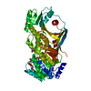
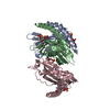
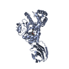
| Title | X-ray crystallographic structure of (6-4)photolyase from Drosophila melanogaster at room temperature | ||||||
 Components Components | RE11660p | ||||||
 Keywords Keywords |  LYASE / Flavoprotein / (6-4)photolyase LYASE / Flavoprotein / (6-4)photolyase | ||||||
| Function / homology |  Function and homology informationdeoxyribodipyrimidine photo-lyase activity / entrainment of circadian clock by photoperiod / FAD binding / circadian regulation of gene expression / DNA binding / nucleus / cytoplasm Function and homology informationdeoxyribodipyrimidine photo-lyase activity / entrainment of circadian clock by photoperiod / FAD binding / circadian regulation of gene expression / DNA binding / nucleus / cytoplasmSimilarity search - Function | ||||||
| Biological species |  Drosophila melanogaster (fruit fly) Drosophila melanogaster (fruit fly) | ||||||
| Method | X-RAY DIFFRACTION / FREE ELECTRON LASER / MOLECULAR REPLACEMENT / Resolution: 2.27 Å | ||||||
 Authors Authors | Cellini, A. / Wahlgren, W.Y. / Henry, L. / Westenhoff, S. / Pandey, S. | ||||||
| Funding support |  Sweden, 1items Sweden, 1items
| ||||||
 Citation Citation | Journal: Acta Crystallogr D Struct Biol / Year: 2021 Title: The three-dimensional structure of Drosophila melanogaster (6-4) photolyase at room temperature. Authors: Cellini, A. / Yuan Wahlgren, W. / Henry, L. / Pandey, S. / Ghosh, S. / Castillon, L. / Claesson, E. / Takala, H. / Kubel, J. / Nimmrich, A. / Kuznetsova, V. / Nango, E. / Iwata, S. / Owada, ...Authors: Cellini, A. / Yuan Wahlgren, W. / Henry, L. / Pandey, S. / Ghosh, S. / Castillon, L. / Claesson, E. / Takala, H. / Kubel, J. / Nimmrich, A. / Kuznetsova, V. / Nango, E. / Iwata, S. / Owada, S. / Stojkovic, E.A. / Schmidt, M. / Ihalainen, J.A. / Westenhoff, S. | ||||||
| History |
|
- Structure visualization
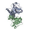
| Structure viewer | Molecule: MolmilJmol/JSmol |
|---|
- Downloads & links
-Download
| PDBx/mmCIF format | 7azt.cif.gz | 136 KB | Display | PDBx/mmCIF format |
|---|---|---|---|---|
| PDB format | pdb7azt.ent.gz | 88 KB | Display | PDB format |
| PDBx/mmJSON format | 7azt.json.gz | Tree view | PDBx/mmJSON format | |
| Others |  Other downloads Other downloads |
-Validation report
| Arichive directory | https://data.pdbj.org/pub/pdb/validation_reports/az/7aztftp://data.pdbj.org/pub/pdb/validation_reports/az/7azt | HTTPS FTP |
|---|
-Related structure data
| Related structure data | 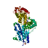 7ayvSC S: Starting model for refinement C: citing same article ( |
|---|---|
| Similar structure data |


-Links
 PDBj
PDBj
- Assembly
| Deposited unit | 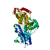
| ||||||||||||
|---|---|---|---|---|---|---|---|---|---|---|---|---|---|
| 1 |
| ||||||||||||
| Unit cell |
|
-Components
| #1: Protein | Mass: 58310.973 Da / Num. of mol.: 1 Source method: isolated from a genetically manipulated source Source: (gene. exp.) Drosophila melanogaster (fruit fly) / Gene: phr6-4, CG2488 / Production host:  Escherichia coli K-12 (bacteria) / References: UniProt: Q8SXK5 Escherichia coli K-12 (bacteria) / References: UniProt: Q8SXK5 |
|---|---|
| #2: Chemical | ChemComp-FAD / Flavin adenine dinucleotide  Mass: 785.550 Da / Num. of mol.: 1 / Source method: obtained synthetically / Formula: C27H33N9O15P2 / Feature type: SUBJECT OF INVESTIGATION / Comment: FAD*YM Mass: 785.550 Da / Num. of mol.: 1 / Source method: obtained synthetically / Formula: C27H33N9O15P2 / Feature type: SUBJECT OF INVESTIGATION / Comment: FAD*YM |
| Has ligand of interest | Y |
-Experimental details
-Experiment
| Experiment | Method: X-RAY DIFFRACTION / Number of used crystals: 1 |
|---|
- Sample preparation
| Crystal | Density Matthews: 2.81 Å3/Da / Density % sol: 56.2 % |
|---|---|
| Crystal grow | Temperature: 277.15 K / Method: batch mode / pH: 6.5 Details: 0.2 M lithium sulftate, 0.1 M bis-tris (pH=6.5), 25% PEG3350, 4% polypropyle glycol P400 |
-Data collection
| Diffraction | Mean temperature: 298 K / Serial crystal experiment: Y |
|---|---|
| Diffraction source | Source: FREE ELECTRON LASER / Site: SACLA  / Beamline: BL3 / Wavelength: 1.7712 Å / Beamline: BL3 / Wavelength: 1.7712 Å |
| Detector | Type: MPCCD / Detector: CCD / Date: May 19, 2019 |
| Radiation | Protocol: SINGLE WAVELENGTH / Monochromatic (M) / Laue (L): M / Scattering type: x-ray |
| Radiation wavelength | Wavelength: 1.7712 Å / Relative weight: 1 |
| Reflection | Resolution: 2.27→42.61 Å / Num. obs: 33953 / % possible obs: 100 % / Redundancy: 106 % / Biso Wilson estimate: 90.69 Å2 / CC1/2: 0.97 / Net I/σ(I): 4.93 |
| Reflection shell | Resolution: 2.27→2.351 Å / Num. unique obs: 1352 / CC1/2: 0.32 |
| Serial crystallography sample delivery | Method: injection |
- Processing
| Software |
| ||||||||||||||||||||||||||||||||||||||||||||||||||||||||||||||||||||||||||||||||||||
|---|---|---|---|---|---|---|---|---|---|---|---|---|---|---|---|---|---|---|---|---|---|---|---|---|---|---|---|---|---|---|---|---|---|---|---|---|---|---|---|---|---|---|---|---|---|---|---|---|---|---|---|---|---|---|---|---|---|---|---|---|---|---|---|---|---|---|---|---|---|---|---|---|---|---|---|---|---|---|---|---|---|---|---|---|---|
| Refinement | Method to determine structure: MOLECULAR REPLACEMENT Starting model: 7AYV Resolution: 2.27→42.61 Å / SU ML: 0.3689 / Cross valid method: FREE R-VALUE / σ(F): 1.34 / Phase error: 28.0904 Stereochemistry target values: GeoStd + Monomer Library + CDL v1.2
| ||||||||||||||||||||||||||||||||||||||||||||||||||||||||||||||||||||||||||||||||||||
| Solvent computation | Shrinkage radii: 0.9 Å / VDW probe radii: 1.11 Å / Solvent model: FLAT BULK SOLVENT MODEL | ||||||||||||||||||||||||||||||||||||||||||||||||||||||||||||||||||||||||||||||||||||
| Displacement parameters | Biso mean: 93.13 Å2 | ||||||||||||||||||||||||||||||||||||||||||||||||||||||||||||||||||||||||||||||||||||
| Refinement step | Cycle: LAST / Resolution: 2.27→42.61 Å
| ||||||||||||||||||||||||||||||||||||||||||||||||||||||||||||||||||||||||||||||||||||
| Refine LS restraints |
| ||||||||||||||||||||||||||||||||||||||||||||||||||||||||||||||||||||||||||||||||||||
| LS refinement shell |
|
