 Movie
Movie Controller

[English] 日本語
 Yorodumi
- PDB-7ayv: X-ray crystallographic structure of (6-4)photolyase from Drosophi... -
+ Open data

- Basic information
| Entry | Database: PDB / ID: 7ayv | ||||||
|---|---|---|---|---|---|---|---|
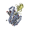
| Title | X-ray crystallographic structure of (6-4)photolyase from Drosophila melanogaster at cryogenic temperature | ||||||
 Components Components | RE11660p | ||||||
 Keywords Keywords |  LYASE / Flavoprotein / (6-4)photolyase LYASE / Flavoprotein / (6-4)photolyase | ||||||
| Function / homology |  Function and homology informationdeoxyribodipyrimidine photo-lyase activity / entrainment of circadian clock by photoperiod / FAD binding / circadian regulation of gene expression / DNA binding / nucleus / cytoplasm Function and homology informationdeoxyribodipyrimidine photo-lyase activity / entrainment of circadian clock by photoperiod / FAD binding / circadian regulation of gene expression / DNA binding / nucleus / cytoplasmSimilarity search - Function | ||||||
| Biological species |  Drosophila melanogaster (fruit fly) Drosophila melanogaster (fruit fly) | ||||||
| Method | X-RAY DIFFRACTION / SYNCHROTRON / MOLECULAR REPLACEMENT / Resolution: 1.79 Å | ||||||
 Authors Authors | Cellini, A. / Wahlgren, W.Y. / Henry, L. / Westenhoff, S. | ||||||
| Funding support |  Sweden, 1items Sweden, 1items
| ||||||
 Citation Citation | Journal: Acta Crystallogr D Struct Biol / Year: 2021 Title: The three-dimensional structure of Drosophila melanogaster (6-4) photolyase at room temperature. Authors: Cellini, A. / Yuan Wahlgren, W. / Henry, L. / Pandey, S. / Ghosh, S. / Castillon, L. / Claesson, E. / Takala, H. / Kubel, J. / Nimmrich, A. / Kuznetsova, V. / Nango, E. / Iwata, S. / Owada, ...Authors: Cellini, A. / Yuan Wahlgren, W. / Henry, L. / Pandey, S. / Ghosh, S. / Castillon, L. / Claesson, E. / Takala, H. / Kubel, J. / Nimmrich, A. / Kuznetsova, V. / Nango, E. / Iwata, S. / Owada, S. / Stojkovic, E.A. / Schmidt, M. / Ihalainen, J.A. / Westenhoff, S. | ||||||
| History |
|
- Structure visualization
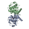
| Structure viewer | Molecule: MolmilJmol/JSmol |
|---|
- Downloads & links
-Download
| PDBx/mmCIF format | 7ayv.cif.gz | 209.7 KB | Display | PDBx/mmCIF format |
|---|---|---|---|---|
| PDB format | pdb7ayv.ent.gz | 134.5 KB | Display | PDB format |
| PDBx/mmJSON format | 7ayv.json.gz | Tree view | PDBx/mmJSON format | |
| Others |  Other downloads Other downloads |
-Validation report
| Arichive directory | https://data.pdbj.org/pub/pdb/validation_reports/ay/7ayvftp://data.pdbj.org/pub/pdb/validation_reports/ay/7ayv | HTTPS FTP |
|---|
-Related structure data
| Related structure data |  7aztC 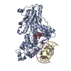 3cvyS S: Starting model for refinement C: citing same article ( |
|---|---|
| Similar structure data |


-Links
 PDBj
PDBj
- Assembly
| Deposited unit | 
| ||||||||||||
|---|---|---|---|---|---|---|---|---|---|---|---|---|---|
| 1 |
| ||||||||||||
| Unit cell |
|
-Components
| #1: Protein | Mass: 58313.043 Da / Num. of mol.: 1 Source method: isolated from a genetically manipulated source Source: (gene. exp.) Drosophila melanogaster (fruit fly) / Gene: phr6-4, CG2488 / Production host:  Escherichia coli K-12 (bacteria) / References: UniProt: Q8SXK5 Escherichia coli K-12 (bacteria) / References: UniProt: Q8SXK5 | ||||
|---|---|---|---|---|---|
| #2: Chemical | ChemComp-GOL / Glycerol  Mass: 92.094 Da / Num. of mol.: 1 / Source method: obtained synthetically / Formula: C3H8O3 Mass: 92.094 Da / Num. of mol.: 1 / Source method: obtained synthetically / Formula: C3H8O3 | ||||
| #3: Chemical | ChemComp-FAD / Flavin adenine dinucleotide  Mass: 785.550 Da / Num. of mol.: 1 / Source method: obtained synthetically / Formula: C27H33N9O15P2 / Feature type: SUBJECT OF INVESTIGATION / Comment: FAD*YM Mass: 785.550 Da / Num. of mol.: 1 / Source method: obtained synthetically / Formula: C27H33N9O15P2 / Feature type: SUBJECT OF INVESTIGATION / Comment: FAD*YM | ||||
| #4: Chemical | ChemComp-SO4 / Sulfate  Mass: 96.063 Da / Num. of mol.: 5 / Source method: obtained synthetically / Formula: SO4 Mass: 96.063 Da / Num. of mol.: 5 / Source method: obtained synthetically / Formula: SO4#5: Water | ChemComp-HOH / | Water Mass: 18.015 Da / Num. of mol.: 253 / Source method: isolated from a natural source / Formula: H2O Mass: 18.015 Da / Num. of mol.: 253 / Source method: isolated from a natural source / Formula: H2OHas ligand of interest | Y | |
-Experimental details
-Experiment
| Experiment | Method: X-RAY DIFFRACTION / Number of used crystals: 1 |
|---|
- Sample preparation
| Crystal | Density Matthews: 2.6 Å3/Da / Density % sol: 52.75 % |
|---|---|
| Crystal grow | Temperature: 293.15 K / Method: vapor diffusion, hanging drop / pH: 6.5 Details: 0.2 M Lithium sulphate, 0.1 M Bis-Tris(pH=6.5), 25%PEG3350, 4% polypropylene glycol p 400 |
-Data collection
| Diffraction | Mean temperature: 100 K / Serial crystal experiment: N |
|---|---|
| Diffraction source | Source: SYNCHROTRON / Site: MAX IV / Beamline: BioMAX / Wavelength: 0.9184 Å |
| Detector | Type: DECTRIS EIGER X 16M / Detector: PIXEL / Date: Mar 29, 2019 |
| Radiation | Protocol: SINGLE WAVELENGTH / Monochromatic (M) / Laue (L): M / Scattering type: x-ray |
| Radiation wavelength | Wavelength: 0.9184 Å / Relative weight: 1 |
| Reflection | Resolution: 1.79→29.42 Å / Num. obs: 58656 / % possible obs: 99.2 % / Redundancy: 12.7 % / Biso Wilson estimate: 25.32 Å2 / CC1/2: 0.99 / Net I/σ(I): 16.9 |
| Reflection shell | Resolution: 1.79→1.854 Å / Num. unique obs: 3836 / CC1/2: 0.83 |
- Processing
| Software |
| |||||||||||||||||||||||||||||||||||||||||||||||||||||||||||||||||||||||||||||||||||||||||||||||||||||||||
|---|---|---|---|---|---|---|---|---|---|---|---|---|---|---|---|---|---|---|---|---|---|---|---|---|---|---|---|---|---|---|---|---|---|---|---|---|---|---|---|---|---|---|---|---|---|---|---|---|---|---|---|---|---|---|---|---|---|---|---|---|---|---|---|---|---|---|---|---|---|---|---|---|---|---|---|---|---|---|---|---|---|---|---|---|---|---|---|---|---|---|---|---|---|---|---|---|---|---|---|---|---|---|---|---|---|---|
| Refinement | Method to determine structure: MOLECULAR REPLACEMENT Starting model: 3cvy Resolution: 1.79→29.42 Å / SU ML: 0.2196 / Cross valid method: FREE R-VALUE / σ(F): 1.34 / Phase error: 19.858 Stereochemistry target values: GeoStd + Monomer Library + CDL v1.2
| |||||||||||||||||||||||||||||||||||||||||||||||||||||||||||||||||||||||||||||||||||||||||||||||||||||||||
| Solvent computation | Shrinkage radii: 0.9 Å / VDW probe radii: 1.11 Å / Solvent model: FLAT BULK SOLVENT MODEL | |||||||||||||||||||||||||||||||||||||||||||||||||||||||||||||||||||||||||||||||||||||||||||||||||||||||||
| Displacement parameters | Biso mean: 26.96 Å2 | |||||||||||||||||||||||||||||||||||||||||||||||||||||||||||||||||||||||||||||||||||||||||||||||||||||||||
| Refinement step | Cycle: LAST / Resolution: 1.79→29.42 Å
| |||||||||||||||||||||||||||||||||||||||||||||||||||||||||||||||||||||||||||||||||||||||||||||||||||||||||
| Refine LS restraints |
| |||||||||||||||||||||||||||||||||||||||||||||||||||||||||||||||||||||||||||||||||||||||||||||||||||||||||
| LS refinement shell |
| |||||||||||||||||||||||||||||||||||||||||||||||||||||||||||||||||||||||||||||||||||||||||||||||||||||||||
| Refinement TLS params. | Method: refined / Refine-ID: X-RAY DIFFRACTION
| |||||||||||||||||||||||||||||||||||||||||||||||||||||||||||||||||||||||||||||||||||||||||||||||||||||||||
| Refinement TLS group |
|
