 Movie
Movie Controller

[English] 日本語
 Yorodumi
- PDB-7ap1: Klebsiella pneumoniae Seryl-tRNA synthetase in Complex with Compo... -
+ Open data

- Basic information
| Entry | Database: PDB / ID: 7ap1 | ||||||||||||
|---|---|---|---|---|---|---|---|---|---|---|---|---|---|
| Title | Klebsiella pneumoniae Seryl-tRNA synthetase in Complex with Compound SerS7HMDDA | ||||||||||||
 Components Components | Serine-tRNA ligase Serine—tRNA ligase Serine—tRNA ligase | ||||||||||||
 Keywords Keywords | LIGASE / protein-inhibitor complex / coiled-coil / tRNA synthetase | ||||||||||||
| Function / homology |  Function and homology information Function and homology informationselenocysteine biosynthetic process / serine-tRNA ligase / serine-tRNA ligase activity / seryl-tRNA aminoacylation / ATP binding / cytoplasmSimilarity search - Function | ||||||||||||
| Biological species |  Klebsiella pneumoniae (bacteria) Klebsiella pneumoniae (bacteria) | ||||||||||||
| Method | X-RAY DIFFRACTION / SYNCHROTRON / MOLECULAR REPLACEMENT / Resolution: 2.18 Å | ||||||||||||
 Authors Authors | Pang, L. / De Graef, S. / Strelkov, S.V. / Weeks, S.D. | ||||||||||||
| Funding support |  Belgium, 3items Belgium, 3items
| ||||||||||||
 Citation Citation | Journal: Molecules / Year: 2020 Title: Synthesis and Biological Evaluation of 1,3-Dideazapurine-Like 7-Amino-5-Hydroxymethyl-Benzimidazole Ribonucleoside Analogues as Aminoacyl-tRNA Synthetase Inhibitors. Authors: Zhang, B. / Pang, L. / Nautiyal, M. / De Graef, S. / Gadakh, B. / Lescrinier, E. / Rozenski, J. / Strelkov, S.V. / Weeks, S.D. / Van Aerschot, A. | ||||||||||||
| History |
|
- Structure visualization
| Structure viewer | Molecule: MolmilJmol/JSmol |
|---|
- Downloads & links
-Download
| PDBx/mmCIF format | 7ap1.cif.gz | 184.5 KB | Display | PDBx/mmCIF format |
|---|---|---|---|---|
| PDB format | pdb7ap1.ent.gz | 145.6 KB | Display | PDB format |
| PDBx/mmJSON format | 7ap1.json.gz | Tree view | PDBx/mmJSON format | |
| Others |  Other downloads Other downloads |
-Validation report
| Arichive directory | https://data.pdbj.org/pub/pdb/validation_reports/ap/7ap1ftp://data.pdbj.org/pub/pdb/validation_reports/ap/7ap1 | HTTPS FTP |
|---|
-Related structure data
| Related structure data | 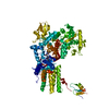 7ap2C 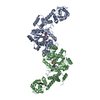 7ap3C  7ap4C  6h9xS C: citing same article ( S: Starting model for refinement |
|---|---|
| Similar structure data |


-Links
 PDBj
PDBj
- Assembly
| Deposited unit | 
| ||||||||
|---|---|---|---|---|---|---|---|---|---|
| 1 | 
| ||||||||
| Unit cell |
|
-Components
| #1: Protein | Serine—tRNA ligase / Seryl-tRNA synthetase / SerRS / Seryl-tRNA(Ser/Sec) synthetase Mass: 48669.180 Da / Num. of mol.: 1 / Fragment: Seryl-tRNA synthetase Source method: isolated from a genetically manipulated source Source: (gene. exp.) Klebsiella pneumoniae (bacteria)Gene: serS, AGG09_05015, B1727_25560, B4U21_07300, B4U22_08075, BB785_18280, BL124_0015080, BN49_2024, C3483_17160, C3F39_05595, C9J88_18700, CPT10_05250, CSC88_02630, CWQ24_18365, PMK1_03261, SM57_00973 Plasmid: pETRUK / Production host: Escherichia coli BL21(DE3) (bacteria) / Strain (production host): Rosetta 2 (DE3) pLysSReferences: UniProt: W9BNU9, UniProt: A6T6Z0*PLUS, serine-tRNA ligase | ||||||
|---|---|---|---|---|---|---|---|
| #2: Chemical | ChemComp-RUZ / [(  Mass: 461.447 Da / Num. of mol.: 1 / Source method: obtained synthetically / Formula: C16H23N5O9S / Feature type: SUBJECT OF INVESTIGATION Mass: 461.447 Da / Num. of mol.: 1 / Source method: obtained synthetically / Formula: C16H23N5O9S / Feature type: SUBJECT OF INVESTIGATION | ||||||
| #3: Chemical | Ethylene glycol  Mass: 62.068 Da / Num. of mol.: 3 / Source method: obtained synthetically / Formula: C2H6O2 Mass: 62.068 Da / Num. of mol.: 3 / Source method: obtained synthetically / Formula: C2H6O2#4: Chemical |   Mass: 40.078 Da / Num. of mol.: 3 / Source method: obtained synthetically / Formula: Ca Mass: 40.078 Da / Num. of mol.: 3 / Source method: obtained synthetically / Formula: Ca#5: Water | ChemComp-HOH / | Water Mass: 18.015 Da / Num. of mol.: 118 / Source method: isolated from a natural source / Formula: H2O Mass: 18.015 Da / Num. of mol.: 118 / Source method: isolated from a natural source / Formula: H2OHas ligand of interest | Y | |
-Experimental details
-Experiment
| Experiment | Method: X-RAY DIFFRACTION / Number of used crystals: 1 |
|---|
- Sample preparation
| Crystal | Density Matthews: 4.23 Å3/Da / Density % sol: 70.94 % / Mosaicity: 0.09 ° |
|---|---|
| Crystal grow | Temperature: 293 K / Method: vapor diffusion, hanging drop / pH: 6.5 Details: Holo protein at 10 mg/mL in 10 mM Tris pH 7, 100 mM NaCl, 5 mM DTT was mixed with an equal volume of 7.5% w/v PEG 8000, 50 mM CaCl2, 20% v/v Ethylene glycol, 100 mM Morpheus buffer system 1 ...Details: Holo protein at 10 mg/mL in 10 mM Tris pH 7, 100 mM NaCl, 5 mM DTT was mixed with an equal volume of 7.5% w/v PEG 8000, 50 mM CaCl2, 20% v/v Ethylene glycol, 100 mM Morpheus buffer system 1 (MES/Imidazole) pH 6.5. Suitable crystals were soaked with 5 mM synthesized compound in the same crystallization solution but containing a 10% w/v PEG 8000 concentration. |
-Data collection
| Diffraction | Mean temperature: 100 K / Serial crystal experiment: N | ||||||||||||||||||||||||||||||
|---|---|---|---|---|---|---|---|---|---|---|---|---|---|---|---|---|---|---|---|---|---|---|---|---|---|---|---|---|---|---|---|
| Diffraction source | Source: SYNCHROTRON / Site: SOLEIL  / Beamline: PROXIMA 1 / Wavelength: 0.99187 Å / Beamline: PROXIMA 1 / Wavelength: 0.99187 Å | ||||||||||||||||||||||||||||||
| Detector | Type: DECTRIS PILATUS 6M / Detector: PIXEL / Date: Oct 1, 2017 | ||||||||||||||||||||||||||||||
| Radiation | Monochromator: Si(111) / Protocol: SINGLE WAVELENGTH / Monochromatic (M) / Laue (L): M / Scattering type: x-ray | ||||||||||||||||||||||||||||||
| Radiation wavelength | Wavelength: 0.99187 Å / Relative weight: 1 | ||||||||||||||||||||||||||||||
| Reflection | Resolution: 1.91→19.96 Å / Num. obs: 65882 / % possible obs: 99.8 % / Redundancy: 12.2 % / Biso Wilson estimate: 53.83 Å2 / CC1/2: 0.999 / Rmerge(I) obs: 0.105 / Rpim(I) all: 0.032 / Rrim(I) all: 0.11 / Net I/σ(I): 10.7 / Num. measured all: 805160 / Scaling rejects: 1 | ||||||||||||||||||||||||||||||
| Reflection shell | Diffraction-ID: 1
|
- Processing
| Software |
| ||||||||||||||||||||||||||||||||||||||||||||||||||||||||||||||||||||||||||||||||||||||||||||||||||||||||||||
|---|---|---|---|---|---|---|---|---|---|---|---|---|---|---|---|---|---|---|---|---|---|---|---|---|---|---|---|---|---|---|---|---|---|---|---|---|---|---|---|---|---|---|---|---|---|---|---|---|---|---|---|---|---|---|---|---|---|---|---|---|---|---|---|---|---|---|---|---|---|---|---|---|---|---|---|---|---|---|---|---|---|---|---|---|---|---|---|---|---|---|---|---|---|---|---|---|---|---|---|---|---|---|---|---|---|---|---|---|---|
| Refinement | Method to determine structure: MOLECULAR REPLACEMENT Starting model: 6H9X Resolution: 2.18→19.96 Å / Cor.coef. Fo:Fc: 0.933 / Cor.coef. Fo:Fc free: 0.928 / SU R Cruickshank DPI: 0.148 / Cross valid method: THROUGHOUT / σ(F): 0 / SU R Blow DPI: 0.149 / SU Rfree Blow DPI: 0.138 / SU Rfree Cruickshank DPI: 0.138
| ||||||||||||||||||||||||||||||||||||||||||||||||||||||||||||||||||||||||||||||||||||||||||||||||||||||||||||
| Displacement parameters | Biso max: 128.26 Å2 / Biso mean: 61.19 Å2 / Biso min: 40.09 Å2
| ||||||||||||||||||||||||||||||||||||||||||||||||||||||||||||||||||||||||||||||||||||||||||||||||||||||||||||
| Refine analyze | Luzzati coordinate error obs: 0.29 Å | ||||||||||||||||||||||||||||||||||||||||||||||||||||||||||||||||||||||||||||||||||||||||||||||||||||||||||||
| Refinement step | Cycle: final / Resolution: 2.18→19.96 Å
| ||||||||||||||||||||||||||||||||||||||||||||||||||||||||||||||||||||||||||||||||||||||||||||||||||||||||||||
| Refine LS restraints |
| ||||||||||||||||||||||||||||||||||||||||||||||||||||||||||||||||||||||||||||||||||||||||||||||||||||||||||||
| LS refinement shell | Resolution: 2.18→2.19 Å / Rfactor Rfree error: 0 / Total num. of bins used: 51
| ||||||||||||||||||||||||||||||||||||||||||||||||||||||||||||||||||||||||||||||||||||||||||||||||||||||||||||
| Refinement TLS params. | Method: refined / Origin x: -12.5785 Å / Origin y: -32.1722 Å / Origin z: -9.7281 Å
| ||||||||||||||||||||||||||||||||||||||||||||||||||||||||||||||||||||||||||||||||||||||||||||||||||||||||||||
| Refinement TLS group | Selection details: { A|* } |
