 Movie
Movie Controller

[English] 日本語
 Yorodumi
- PDB-6hhz: Klebsiella pneumoniae Seryl-tRNA Synthetase in Complex with 5'-O-... -
+ Open data

- Basic information
| Entry | Database: PDB / ID: 6hhz | ||||||||||||
|---|---|---|---|---|---|---|---|---|---|---|---|---|---|
| Title | Klebsiella pneumoniae Seryl-tRNA Synthetase in Complex with 5'-O-(N-(L-seryl)-sulfamoyl)cytidine | ||||||||||||
 Components Components | Serine--tRNA ligase | ||||||||||||
 Keywords Keywords |  LIGASE / Coil-coil / Beta barrel / tRNA synthetase / Inhibitor / Complex LIGASE / Coil-coil / Beta barrel / tRNA synthetase / Inhibitor / Complex | ||||||||||||
| Function / homology |  Function and homology information Function and homology informationselenocysteine biosynthetic process / seryl-tRNA aminoacylation / serine-tRNA ligase / serine-tRNA ligase activity / ATP binding / cytoplasmSimilarity search - Function | ||||||||||||
| Biological species |  Klebsiella pneumoniae (bacteria) Klebsiella pneumoniae (bacteria) | ||||||||||||
| Method | X-RAY DIFFRACTION / SYNCHROTRON / MOLECULAR REPLACEMENT / Resolution: 2.15 Å | ||||||||||||
 Authors Authors | Pang, L. / De Graef, S. / Weeks, S.D. / Strelkov, S.V. | ||||||||||||
| Funding support |  Belgium, 3items Belgium, 3items
| ||||||||||||
 Citation Citation | Journal: Acs Chem.Biol. / Year: 2020 Title: Structural Insights into the Binding of Natural Pyrimidine-Based Inhibitors of Class II Aminoacyl-tRNA Synthetases. Authors: Pang, L. / Nautiyal, M. / De Graef, S. / Gadakh, B. / Zorzini, V. / Economou, A. / Strelkov, S.V. / Van Aerschot, A. / Weeks, S.D. | ||||||||||||
| History |
|
- Structure visualization
| Structure viewer | Molecule: MolmilJmol/JSmol |
|---|
- Downloads & links
-Download
| PDBx/mmCIF format | 6hhz.cif.gz | 187.7 KB | Display | PDBx/mmCIF format |
|---|---|---|---|---|
| PDB format | pdb6hhz.ent.gz | 146.8 KB | Display | PDB format |
| PDBx/mmJSON format | 6hhz.json.gz | Tree view | PDBx/mmJSON format | |
| Others |  Other downloads Other downloads |
-Validation report
| Arichive directory | https://data.pdbj.org/pub/pdb/validation_reports/hh/6hhzftp://data.pdbj.org/pub/pdb/validation_reports/hh/6hhz | HTTPS FTP |
|---|
-Related structure data
| Related structure data |  6hdzC  6he1C  6he3C  6hhvC  6hhwC  6hhxC  6hhyC  6hi0C  6rltC 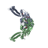 6rluC 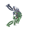 6rlvC  6s30C  6sjcC  2dq3S C: citing same article ( S: Starting model for refinement |
|---|---|
| Similar structure data |


-Links
 PDBj
PDBj
- Assembly
| Deposited unit | 
| ||||||||
|---|---|---|---|---|---|---|---|---|---|
| 1 | 
| ||||||||
| Unit cell |
| ||||||||
| Components on special symmetry positions |
|
-Components
-Protein , 1 types, 1 molecules A
| #1: Protein | Mass: 48669.180 Da / Num. of mol.: 1 / Fragment: Seryl-tRNA synthetase Source method: isolated from a genetically manipulated source Source: (gene. exp.) Klebsiella pneumoniae (bacteria) / Gene: serS, KPN78578_09000, KPN_00925 / Plasmid: pETRUK / Production host: Escherichia coli BL21(DE3) (bacteria) / Variant (production host): Rosetta 2 pLysS / References: UniProt: A6T6Z0, serine-tRNA ligase |
|---|
-Non-polymers , 6 types, 190 molecules 


| #2: Chemical | Ethylene glycol Mass: 62.068 Da / Num. of mol.: 3 / Source method: obtained synthetically / Formula: C2H6O2 Mass: 62.068 Da / Num. of mol.: 3 / Source method: obtained synthetically / Formula: C2H6O2#3: Chemical | ChemComp-FZQ / |  Mass: 409.372 Da / Num. of mol.: 1 / Source method: obtained synthetically / Formula: C12H19N5O9S / Feature type: SUBJECT OF INVESTIGATION Mass: 409.372 Da / Num. of mol.: 1 / Source method: obtained synthetically / Formula: C12H19N5O9S / Feature type: SUBJECT OF INVESTIGATION#4: Chemical |  Mass: 40.078 Da / Num. of mol.: 3 / Source method: obtained synthetically / Formula: Ca Mass: 40.078 Da / Num. of mol.: 3 / Source method: obtained synthetically / Formula: Ca#5: Chemical | ChemComp-CL / | Chloride Mass: 35.453 Da / Num. of mol.: 1 / Source method: obtained synthetically / Formula: Cl Mass: 35.453 Da / Num. of mol.: 1 / Source method: obtained synthetically / Formula: Cl#6: Chemical | ChemComp-1PE / | Polyethylene glycol Mass: 238.278 Da / Num. of mol.: 1 / Source method: obtained synthetically / Formula: C10H22O6 / Comment: precipitant*YM Mass: 238.278 Da / Num. of mol.: 1 / Source method: obtained synthetically / Formula: C10H22O6 / Comment: precipitant*YM#7: Water | ChemComp-HOH / | WaterMass: 18.015 Da / Num. of mol.: 181 / Source method: isolated from a natural source / Formula: H2O |
|---|
-Experimental details
-Experiment
| Experiment | Method: X-RAY DIFFRACTION / Number of used crystals: 1 |
|---|
- Sample preparation
| Crystal | Density Matthews: 4.09 Å3/Da / Density % sol: 69.94 % |
|---|---|
| Crystal grow | Temperature: 293 K / Method: vapor diffusion, hanging drop / pH: 6.5 Details: Holo protein, concentrated to 10 mg/mL in 10 mM Tris pH 7, 100 mM NaCl, 5 mM DTT, was mixed with an equal volume of 7.5% w/v PEG 8000, 50 mM CaCl2, 20% v/v ethylene glycol, 100 mM Morpheus ...Details: Holo protein, concentrated to 10 mg/mL in 10 mM Tris pH 7, 100 mM NaCl, 5 mM DTT, was mixed with an equal volume of 7.5% w/v PEG 8000, 50 mM CaCl2, 20% v/v ethylene glycol, 100 mM Morpheus buffer system 1 (MES/Imidazole) pH 6.5. Suitable crystals were soaked with 2 mM compound in a similar crystallization solution containing a 10% w/v PEG 8000. |
-Data collection
| Diffraction | Mean temperature: 100 K | |||||||||||||||||||||
|---|---|---|---|---|---|---|---|---|---|---|---|---|---|---|---|---|---|---|---|---|---|---|
| Diffraction source | Source: SYNCHROTRON / Site: ESRF  / Beamline: ID23-1 / Wavelength: 0.972422 Å / Beamline: ID23-1 / Wavelength: 0.972422 Å | |||||||||||||||||||||
| Detector | Type: DECTRIS PILATUS 6M / Detector: PIXEL / Date: Dec 17, 2017 | |||||||||||||||||||||
| Radiation | Protocol: SINGLE WAVELENGTH / Monochromatic (M) / Laue (L): M / Scattering type: x-ray | |||||||||||||||||||||
| Radiation wavelength | Wavelength: 0.972422 Å / Relative weight: 1 | |||||||||||||||||||||
| Reflection | Resolution: 1.93→114.66 Å / Num. obs: 61829 / % possible obs: 99.9 % / Redundancy: 8.5 % / Biso Wilson estimate: 50.8 Å2 / CC1/2: 0.999 / Rmerge(I) obs: 0.094 / Rpim(I) all: 0.034 / Rrim(I) all: 0.1 / Net I/σ(I): 10.7 / Num. measured all: 523095 / Scaling rejects: 1 | |||||||||||||||||||||
| Reflection shell | Diffraction-ID: 1 / % possible all: 99.8
|
- Processing
| Software |
| ||||||||||||||||||||||||||||||||||||||||||||||||||||||||||||||||||||||||||||||||||||||||||||||||||||||||||||
|---|---|---|---|---|---|---|---|---|---|---|---|---|---|---|---|---|---|---|---|---|---|---|---|---|---|---|---|---|---|---|---|---|---|---|---|---|---|---|---|---|---|---|---|---|---|---|---|---|---|---|---|---|---|---|---|---|---|---|---|---|---|---|---|---|---|---|---|---|---|---|---|---|---|---|---|---|---|---|---|---|---|---|---|---|---|---|---|---|---|---|---|---|---|---|---|---|---|---|---|---|---|---|---|---|---|---|---|---|---|
| Refinement | Method to determine structure: MOLECULAR REPLACEMENT Starting model: Model generated from structure 2DQ3 Resolution: 2.15→43 Å / Cor.coef. Fo:Fc: 0.932 / Cor.coef. Fo:Fc free: 0.92 / SU R Cruickshank DPI: 0.146 / Cross valid method: THROUGHOUT / σ(F): 0 / SU R Blow DPI: 0.151 / SU Rfree Blow DPI: 0.139 / SU Rfree Cruickshank DPI: 0.137
| ||||||||||||||||||||||||||||||||||||||||||||||||||||||||||||||||||||||||||||||||||||||||||||||||||||||||||||
| Displacement parameters | Biso max: 158.34 Å2 / Biso mean: 55.1 Å2 / Biso min: 32.14 Å2
| ||||||||||||||||||||||||||||||||||||||||||||||||||||||||||||||||||||||||||||||||||||||||||||||||||||||||||||
| Refine analyze | Luzzati coordinate error obs: 0.27 Å | ||||||||||||||||||||||||||||||||||||||||||||||||||||||||||||||||||||||||||||||||||||||||||||||||||||||||||||
| Refinement step | Cycle: final / Resolution: 2.15→43 Å
| ||||||||||||||||||||||||||||||||||||||||||||||||||||||||||||||||||||||||||||||||||||||||||||||||||||||||||||
| Refine LS restraints |
| ||||||||||||||||||||||||||||||||||||||||||||||||||||||||||||||||||||||||||||||||||||||||||||||||||||||||||||
| LS refinement shell | Resolution: 2.15→2.21 Å / Rfactor Rfree error: 0 / Total num. of bins used: 20
| ||||||||||||||||||||||||||||||||||||||||||||||||||||||||||||||||||||||||||||||||||||||||||||||||||||||||||||
| Refinement TLS params. | Method: refined / Origin x: -11.7862 Å / Origin y: -31.1086 Å / Origin z: -9.6704 Å
| ||||||||||||||||||||||||||||||||||||||||||||||||||||||||||||||||||||||||||||||||||||||||||||||||||||||||||||
| Refinement TLS group | Selection details: {A|*} |
