 Movie
Movie Controller


+ Open data

- Basic information
| Entry | Database: PDB / ID: 7a3f | ||||||
|---|---|---|---|---|---|---|---|
| Title | Crystal structure of apo DPP9 | ||||||
 Components Components | Dipeptidyl peptidase 9 | ||||||
 Keywords Keywords | HYDROLASE / DPP9 / Protease | ||||||
| Function / homology |  Function and homology informationdipeptidyl-peptidase IV / dipeptidyl-peptidase activity / negative regulation of programmed cell death / pyroptotic inflammatory response / cell leading edge / aminopeptidase activity / serine-type peptidase activity / microtubule / proteolysis / identical protein binding ...dipeptidyl-peptidase IV / dipeptidyl-peptidase activity / negative regulation of programmed cell death / pyroptotic inflammatory response / cell leading edge / aminopeptidase activity / serine-type peptidase activity / microtubule / proteolysis / identical protein binding / nucleus / cytosol Function and homology informationdipeptidyl-peptidase IV / dipeptidyl-peptidase activity / negative regulation of programmed cell death / pyroptotic inflammatory response / cell leading edge / aminopeptidase activity / serine-type peptidase activity / microtubule / proteolysis / identical protein binding ...dipeptidyl-peptidase IV / dipeptidyl-peptidase activity / negative regulation of programmed cell death / pyroptotic inflammatory response / cell leading edge / aminopeptidase activity / serine-type peptidase activity / microtubule / proteolysis / identical protein binding / nucleus / cytosolSimilarity search - Function | ||||||
| Biological species |  Homo sapiens (human) Homo sapiens (human) | ||||||
| Method | X-RAY DIFFRACTION / SYNCHROTRON / MOLECULAR REPLACEMENT / Resolution: 2.9 Å | ||||||
 Authors Authors | Ross, B.H. / Huber, R. | ||||||
 Citation Citation | Journal: To Be Published Title: Discovery and Development of 4-Oxo-beta-Lactams as Novel Inhibitors of Dipeptidyl Peptidases 8 and 9 Authors: Fehr, L. / Carvalho, L.A.R. / Ross, B.H. / Lum, K. / Vieira, A.C. / Kiefersauer, R. / Geiss-Friedlander, R. / Kaiser, M. / Rodrigues, T. / Lucas, S.D. / Cravatt, B.F. / Huber, R. / Moreira, R. | ||||||
| History |
|
- Structure visualization
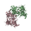
| Structure viewer | Molecule: MolmilJmol/JSmol |
|---|
- Downloads & links
-Download
| PDBx/mmCIF format | 7a3f.cif.gz | 643.8 KB | Display | PDBx/mmCIF format |
|---|---|---|---|---|
| PDB format | pdb7a3f.ent.gz | 516.9 KB | Display | PDB format |
| PDBx/mmJSON format | 7a3f.json.gz | Tree view | PDBx/mmJSON format | |
| Others |  Other downloads Other downloads |
-Validation report
| Arichive directory | https://data.pdbj.org/pub/pdb/validation_reports/a3/7a3fftp://data.pdbj.org/pub/pdb/validation_reports/a3/7a3f | HTTPS FTP |
|---|
-Related structure data
| Related structure data | 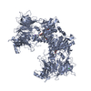 7a3iC 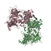 7a3kC 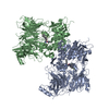 6eorS S: Starting model for refinement C: citing same article ( |
|---|---|
| Similar structure data |


-Links
 PDBj
PDBj
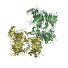
- Assembly
| Deposited unit | 
| ||||||||
|---|---|---|---|---|---|---|---|---|---|
| 1 | 
| ||||||||
| 2 | 
| ||||||||
| Unit cell |
|
-Components
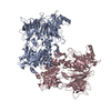
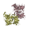
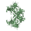
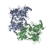
| #1: Protein | / DP9 / Dipeptidyl peptidase IV-related protein 2 / DPRP-2 / Dipeptidyl peptidase IX / DPP IX / ...DP9 / Dipeptidyl peptidase IV-related protein 2 / DPRP-2 / Dipeptidyl peptidase IX / DPP IX / Dipeptidyl peptidase-like protein 9 / DPLP9 Mass: 102627.156 Da / Num. of mol.: 4 Source method: isolated from a genetically manipulated source Source: (gene. exp.) Homo sapiens (human) / Gene: DPP9, DPRP2 / Production host:   Spodoptera frugiperda (fall armyworm) / References: UniProt: Q86TI2, dipeptidyl-peptidase IV Spodoptera frugiperda (fall armyworm) / References: UniProt: Q86TI2, dipeptidyl-peptidase IV#2: Chemical | ChemComp-PO4 / Phosphate  Mass: 94.971 Da / Num. of mol.: 4 / Source method: obtained synthetically / Formula: PO4 / Feature type: SUBJECT OF INVESTIGATION Mass: 94.971 Da / Num. of mol.: 4 / Source method: obtained synthetically / Formula: PO4 / Feature type: SUBJECT OF INVESTIGATION#3: Chemical | Glycerol  Mass: 92.094 Da / Num. of mol.: 2 / Source method: obtained synthetically / Formula: C3H8O3 Mass: 92.094 Da / Num. of mol.: 2 / Source method: obtained synthetically / Formula: C3H8O3#4: Water | ChemComp-HOH / | Water Mass: 18.015 Da / Num. of mol.: 129 / Source method: isolated from a natural source / Formula: H2O Mass: 18.015 Da / Num. of mol.: 129 / Source method: isolated from a natural source / Formula: H2OHas ligand of interest | Y | |
|---|
-Experimental details
-Experiment
| Experiment | Method: X-RAY DIFFRACTION / Number of used crystals: 1 |
|---|
- Sample preparation
| Crystal | Density Matthews: 2.68 Å3/Da / Density % sol: 54.08 % |
|---|---|
| Crystal grow | Temperature: 293 K / Method: vapor diffusion, hanging drop Details: 0.08 M Na-Cacodilate pH 5.25, 0.16 M Ca-Acetate, 30 % Glycerol, 10 % PEG 8K |
-Data collection
| Diffraction | Mean temperature: 100 K / Serial crystal experiment: N | ||||||||||||||||||||||||||||||||||||||||||||||||||||||||||||||||||||||||||||||||||||||||||||||||||||||||||||||||||||||||||||||||||||||||||||||||||||||||||||||||||||||||||||||||||||||||||||||
|---|---|---|---|---|---|---|---|---|---|---|---|---|---|---|---|---|---|---|---|---|---|---|---|---|---|---|---|---|---|---|---|---|---|---|---|---|---|---|---|---|---|---|---|---|---|---|---|---|---|---|---|---|---|---|---|---|---|---|---|---|---|---|---|---|---|---|---|---|---|---|---|---|---|---|---|---|---|---|---|---|---|---|---|---|---|---|---|---|---|---|---|---|---|---|---|---|---|---|---|---|---|---|---|---|---|---|---|---|---|---|---|---|---|---|---|---|---|---|---|---|---|---|---|---|---|---|---|---|---|---|---|---|---|---|---|---|---|---|---|---|---|---|---|---|---|---|---|---|---|---|---|---|---|---|---|---|---|---|---|---|---|---|---|---|---|---|---|---|---|---|---|---|---|---|---|---|---|---|---|---|---|---|---|---|---|---|---|---|---|---|---|
| Diffraction source | Source: SYNCHROTRON / Site: SLS  / Beamline: X10SA / Wavelength: 1 Å / Beamline: X10SA / Wavelength: 1 Å | ||||||||||||||||||||||||||||||||||||||||||||||||||||||||||||||||||||||||||||||||||||||||||||||||||||||||||||||||||||||||||||||||||||||||||||||||||||||||||||||||||||||||||||||||||||||||||||||
| Detector | Type: DECTRIS EIGER X 16M / Detector: PIXEL / Date: Aug 28, 2019 | ||||||||||||||||||||||||||||||||||||||||||||||||||||||||||||||||||||||||||||||||||||||||||||||||||||||||||||||||||||||||||||||||||||||||||||||||||||||||||||||||||||||||||||||||||||||||||||||
| Radiation | Protocol: SINGLE WAVELENGTH / Monochromatic (M) / Laue (L): M / Scattering type: x-ray | ||||||||||||||||||||||||||||||||||||||||||||||||||||||||||||||||||||||||||||||||||||||||||||||||||||||||||||||||||||||||||||||||||||||||||||||||||||||||||||||||||||||||||||||||||||||||||||||
| Radiation wavelength | Wavelength: 1 Å / Relative weight: 1 | ||||||||||||||||||||||||||||||||||||||||||||||||||||||||||||||||||||||||||||||||||||||||||||||||||||||||||||||||||||||||||||||||||||||||||||||||||||||||||||||||||||||||||||||||||||||||||||||
| Reflection | Resolution: 2.9→46.89 Å / Num. obs: 92941 / % possible obs: 98.4 % / Redundancy: 2.235 % / Biso Wilson estimate: 50.051 Å2 / CC1/2: 0.991 / Rmerge(I) obs: 0.087 / Rrim(I) all: 0.115 / Χ2: 0.946 / Net I/σ(I): 7.24 / Num. measured all: 207713 / Scaling rejects: 330 | ||||||||||||||||||||||||||||||||||||||||||||||||||||||||||||||||||||||||||||||||||||||||||||||||||||||||||||||||||||||||||||||||||||||||||||||||||||||||||||||||||||||||||||||||||||||||||||||
| Reflection shell | Diffraction-ID: 1
|
- Processing
| Software |
| ||||||||||||||||||||||||||||||||||||||||||||||||||||||||||||
|---|---|---|---|---|---|---|---|---|---|---|---|---|---|---|---|---|---|---|---|---|---|---|---|---|---|---|---|---|---|---|---|---|---|---|---|---|---|---|---|---|---|---|---|---|---|---|---|---|---|---|---|---|---|---|---|---|---|---|---|---|---|
| Refinement | Method to determine structure: MOLECULAR REPLACEMENT Starting model: 6EOR Resolution: 2.9→46.89 Å / Cor.coef. Fo:Fc: 0.928 / Cor.coef. Fo:Fc free: 0.87 / SU B: 25.629 / SU ML: 0.448 / Cross valid method: THROUGHOUT / σ(F): 0 / ESU R Free: 0.464 Details: HYDROGENS HAVE BEEN ADDED IN THE RIDING POSITIONS U VALUES : REFINED INDIVIDUALLY
| ||||||||||||||||||||||||||||||||||||||||||||||||||||||||||||
| Solvent computation | Ion probe radii: 0.8 Å / Shrinkage radii: 0.8 Å / VDW probe radii: 1.2 Å | ||||||||||||||||||||||||||||||||||||||||||||||||||||||||||||
| Displacement parameters | Biso max: 142.73 Å2 / Biso mean: 59.125 Å2 / Biso min: 9.88 Å2
| ||||||||||||||||||||||||||||||||||||||||||||||||||||||||||||
| Refinement step | Cycle: final / Resolution: 2.9→46.89 Å
| ||||||||||||||||||||||||||||||||||||||||||||||||||||||||||||
| Refine LS restraints |
| ||||||||||||||||||||||||||||||||||||||||||||||||||||||||||||
| LS refinement shell | Resolution: 2.9→2.975 Å / Rfactor Rfree error: 0 / Total num. of bins used: 20
|
