 Movie
Movie Controller

[English] 日本語
 Yorodumi
- PDB-7a2o: Crystal structure of the Fyn SH3 domain L112V-S114N-S115T-E121L-R... -
+ Open data

- Basic information
| Entry | Database: PDB / ID: 7a2o | ||||||
|---|---|---|---|---|---|---|---|
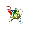
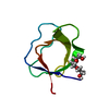
| Title | Crystal structure of the Fyn SH3 domain L112V-S114N-S115T-E121L-R123H mutant at pH 4.5 | ||||||
 Components Components | Tyrosine-protein kinase Fyn | ||||||
 Keywords Keywords |  PROTEIN BINDING / beta barrel / SH3 domain PROTEIN BINDING / beta barrel / SH3 domain | ||||||
| Function / homology |  Function and homology information Function and homology informationresponse to singlet oxygen / Reelin signalling pathway / negative regulation of hydrogen peroxide biosynthetic process / perinuclear endoplasmic reticulum / NTRK2 activates RAC1 / growth factor receptor binding / Activated NTRK2 signals through FYN / heart process / cellular response to L-glutamate / SEMA3A-Plexin repulsion signaling by inhibiting Integrin adhesion ...response to singlet oxygen / Reelin signalling pathway / negative regulation of hydrogen peroxide biosynthetic process / perinuclear endoplasmic reticulum / NTRK2 activates RAC1 / growth factor receptor binding / Activated NTRK2 signals through FYN / heart process / cellular response to L-glutamate / SEMA3A-Plexin repulsion signaling by inhibiting Integrin adhesion / regulation of glutamate receptor signaling pathway / regulation of calcium ion import across plasma membrane / Platelet Adhesion to exposed collagen / G protein-coupled glutamate receptor signaling pathway / CD28 co-stimulation / positive regulation of protein localization to membrane / activated T cell proliferation / CRMPs in Sema3A signaling / positive regulation of cysteine-type endopeptidase activity / FLT3 signaling through SRC family kinases / feeding behavior / Nef and signal transduction / negative regulation of dendritic spine maintenance / type 5 metabotropic glutamate receptor binding / Nephrin family interactions / DCC mediated attractive signaling / dendrite morphogenesis / EPH-Ephrin signaling / CD28 dependent Vav1 pathway / Ephrin signaling / dendritic spine maintenance / Regulation of KIT signaling / tau-protein kinase activity / CTLA4 inhibitory signaling / phospholipase activator activity / leukocyte migration / Fc-gamma receptor signaling pathway involved in phagocytosis / EPHA-mediated growth cone collapse / cellular response to platelet-derived growth factor stimulus / Dectin-2 family / stimulatory C-type lectin receptor signaling pathway / glial cell projection / PECAM1 interactions / CD28 dependent PI3K/Akt signaling / phospholipase binding / response to amyloid-beta / Sema3A PAK dependent Axon repulsion / cellular response to glycine / alpha-tubulin binding / FCGR activation / EPH-ephrin mediated repulsion of cells / positive regulation of protein targeting to membrane / ephrin receptor signaling pathway / Role of LAT2/NTAL/LAB on calcium mobilization / vascular endothelial growth factor receptor signaling pathway / detection of mechanical stimulus involved in sensory perception of pain / negative regulation of oxidative stress-induced intrinsic apoptotic signaling pathway / regulation of peptidyl-tyrosine phosphorylation / forebrain development / positive regulation of tyrosine phosphorylation of STAT protein / negative regulation of inflammatory response to antigenic stimulus / extrinsic component of cytoplasmic side of plasma membrane / Signaling by ERBB2 / negative regulation of protein ubiquitination / GPVI-mediated activation cascade / cellular response to transforming growth factor beta stimulus / EPHB-mediated forward signaling / T cell costimulation / NCAM signaling for neurite out-growth / CD209 (DC-SIGN) signaling / ephrin receptor binding / FCGR3A-mediated IL10 synthesis / Signaling by phosphorylated juxtamembrane, extracellular and kinase domain KIT mutants / Antigen activates B Cell Receptor (BCR) leading to generation of second messengers / learning / actin filament / Regulation of signaling by CBL / Cell surface interactions at the vascular wall / FCGR3A-mediated phagocytosis / cell surface receptor protein tyrosine kinase signaling pathway / axon guidance / non-specific protein-tyrosine kinase / neuron migration / non-membrane spanning protein tyrosine kinase activity / Schaffer collateral - CA1 synapse / protein catabolic process / modulation of chemical synaptic transmission / tau protein binding / Signaling by SCF-KIT / negative regulation of protein catabolic process / VEGFA-VEGFR2 Pathway / positive regulation of neuron projection development / cellular response to hydrogen peroxide / positive regulation of protein localization to nucleus / peptidyl-tyrosine phosphorylation / cellular response to amyloid-beta / Constitutive Signaling by Aberrant PI3K in Cancer / calcium ion transport / Signaling by CSF1 (M-CSF) in myeloid cells / disordered domain specific bindingSimilarity search - Function | ||||||
| Biological species |  Homo sapiens (human) Homo sapiens (human) | ||||||
| Method | X-RAY DIFFRACTION / SYNCHROTRON / MOLECULAR REPLACEMENT / molecular replacement / Resolution: 0.94 Å | ||||||
| Model details | Fyn-SH3-RT chimeric construction | ||||||
 Authors Authors | Camara-Artigas, A. / Plaza-Garrido, M. / Salinas-Garcia, M.C. | ||||||
| Funding support |  Spain, 1items Spain, 1items
| ||||||
 Citation Citation | Journal: To be published Title: Crystal structure of the Fyn SH3 domain L112V-S114N-S115T-E121L-R123H mutant at pH 4.5 Authors: Camara-Artigas, A. / Plaza-Garrido, M. / Salinas-Garcia, M.C. | ||||||
| History |
|
- Structure visualization
| Structure viewer | Molecule: MolmilJmol/JSmol |
|---|
- Downloads & links
-Download
| PDBx/mmCIF format | 7a2o.cif.gz | 53.4 KB | Display | PDBx/mmCIF format |
|---|---|---|---|---|
| PDB format | pdb7a2o.ent.gz | 37.9 KB | Display | PDB format |
| PDBx/mmJSON format | 7a2o.json.gz | Tree view | PDBx/mmJSON format | |
| Others |  Other downloads Other downloads |
-Validation report
| Arichive directory | https://data.pdbj.org/pub/pdb/validation_reports/a2/7a2oftp://data.pdbj.org/pub/pdb/validation_reports/a2/7a2o | HTTPS FTP |
|---|
-Related structure data
| Related structure data |  3ua6S S: Starting model for refinement |
|---|---|
| Similar structure data |


-Links
 PDBj
PDBj


- Assembly
| Deposited unit | 
| ||||||||
|---|---|---|---|---|---|---|---|---|---|
| 1 |
| ||||||||
| Unit cell |
|
-Components
| #1: Protein | Mass: 6787.357 Da / Num. of mol.: 1 / Mutation: L112V S114N S115T E121L R123H Source method: isolated from a genetically manipulated source Source: (gene. exp.) Homo sapiens (human) / Gene: FYN / Plasmid: pHTP1 / Production host:  Escherichia coli BL21(DE3) (bacteria) Escherichia coli BL21(DE3) (bacteria)References: UniProt: P06241, non-specific protein-tyrosine kinase |
|---|---|
| #2: Chemical | ChemComp-PGE / Polyethylene glycol  Mass: 150.173 Da / Num. of mol.: 1 / Source method: obtained synthetically / Formula: C6H14O4 Mass: 150.173 Da / Num. of mol.: 1 / Source method: obtained synthetically / Formula: C6H14O4 |
| #3: Water | ChemComp-HOH / Water Mass: 18.015 Da / Num. of mol.: 85 / Source method: isolated from a natural source / Formula: H2O Mass: 18.015 Da / Num. of mol.: 85 / Source method: isolated from a natural source / Formula: H2O |
| Has ligand of interest | N |
-Experimental details
-Experiment
| Experiment | Method: X-RAY DIFFRACTION / Number of used crystals: 1 |
|---|
- Sample preparation
| Crystal | Density Matthews: 2.06 Å3/Da / Density % sol: 40.2 % / Mosaicity: 0.12 ° |
|---|---|
| Crystal grow | Temperature: 298 K / Method: vapor diffusion, sitting drop / pH: 4.5 Details: 5% PEG 300, 1.5 ammonium sulfate, 0.1M sodium acetate |
-Data collection
| Diffraction | Mean temperature: 100 K / Serial crystal experiment: N | ||||||||||||||||||||||||||||||
|---|---|---|---|---|---|---|---|---|---|---|---|---|---|---|---|---|---|---|---|---|---|---|---|---|---|---|---|---|---|---|---|
| Diffraction source | Source: SYNCHROTRON / Site: ALBA / Beamline: XALOC / Wavelength: 0.97879 Å | ||||||||||||||||||||||||||||||
| Detector | Type: DECTRIS PILATUS 6M / Detector: PIXEL / Date: Dec 2, 2017 | ||||||||||||||||||||||||||||||
| Radiation | Protocol: SINGLE WAVELENGTH / Monochromatic (M) / Laue (L): M / Scattering type: x-ray | ||||||||||||||||||||||||||||||
| Radiation wavelength | Wavelength: 0.97879 Å / Relative weight: 1 | ||||||||||||||||||||||||||||||
| Reflection | Resolution: 0.94→59.66 Å / Num. obs: 69035 / % possible obs: 98.8 % / Redundancy: 5.7 % / CC1/2: 0.997 / Rmerge(I) obs: 0.059 / Rpim(I) all: 0.026 / Rrim(I) all: 0.064 / Net I/σ(I): 16.4 | ||||||||||||||||||||||||||||||
| Reflection shell | Diffraction-ID: 1
|
-Phasing
| Phasing | Method: molecular replacement |
|---|
- Processing
| Software |
| |||||||||||||||||||||||||||||||||||||||||||||||||||||||||||||||||||||||||||||||||||||||||||||||||||||||||||||||||||||||||||||||||||||||||||||||||||||||||||||||||||||||||||||||
|---|---|---|---|---|---|---|---|---|---|---|---|---|---|---|---|---|---|---|---|---|---|---|---|---|---|---|---|---|---|---|---|---|---|---|---|---|---|---|---|---|---|---|---|---|---|---|---|---|---|---|---|---|---|---|---|---|---|---|---|---|---|---|---|---|---|---|---|---|---|---|---|---|---|---|---|---|---|---|---|---|---|---|---|---|---|---|---|---|---|---|---|---|---|---|---|---|---|---|---|---|---|---|---|---|---|---|---|---|---|---|---|---|---|---|---|---|---|---|---|---|---|---|---|---|---|---|---|---|---|---|---|---|---|---|---|---|---|---|---|---|---|---|---|---|---|---|---|---|---|---|---|---|---|---|---|---|---|---|---|---|---|---|---|---|---|---|---|---|---|---|---|---|---|---|---|---|
| Refinement | Method to determine structure: MOLECULAR REPLACEMENT Starting model: 3UA6 Resolution: 0.94→21.86 Å / SU ML: 0.1 / Cross valid method: THROUGHOUT / σ(F): 0.44 / Phase error: 16.89 / Stereochemistry target values: ML
| |||||||||||||||||||||||||||||||||||||||||||||||||||||||||||||||||||||||||||||||||||||||||||||||||||||||||||||||||||||||||||||||||||||||||||||||||||||||||||||||||||||||||||||||
| Solvent computation | Shrinkage radii: 0.9 Å / VDW probe radii: 1.11 Å / Solvent model: FLAT BULK SOLVENT MODEL | |||||||||||||||||||||||||||||||||||||||||||||||||||||||||||||||||||||||||||||||||||||||||||||||||||||||||||||||||||||||||||||||||||||||||||||||||||||||||||||||||||||||||||||||
| Displacement parameters | Biso max: 54.14 Å2 / Biso mean: 14.9456 Å2 / Biso min: 7.14 Å2 | |||||||||||||||||||||||||||||||||||||||||||||||||||||||||||||||||||||||||||||||||||||||||||||||||||||||||||||||||||||||||||||||||||||||||||||||||||||||||||||||||||||||||||||||
| Refinement step | Cycle: final / Resolution: 0.94→21.86 Å
| |||||||||||||||||||||||||||||||||||||||||||||||||||||||||||||||||||||||||||||||||||||||||||||||||||||||||||||||||||||||||||||||||||||||||||||||||||||||||||||||||||||||||||||||
| LS refinement shell | Refine-ID: X-RAY DIFFRACTION / Rfactor Rfree error: 0 / Total num. of bins used: 24
|
