 Movie
Movie Controller

[English] 日本語
 Yorodumi
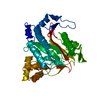
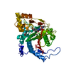
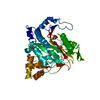
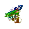
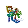
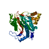
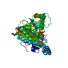
- PDB-7a16: CRYSTAL STRUCTURE OF HUMAN METHIONINE AMINOPEPTIDASE-2 IN COMPLEX... -
+ Open data

- Basic information
| Entry | Database: PDB / ID: 7a16 | ||||||
|---|---|---|---|---|---|---|---|
| Title | CRYSTAL STRUCTURE OF HUMAN METHIONINE AMINOPEPTIDASE-2 IN COMPLEX WITH AN INHIBITOR GSK2229238A (COMPOUND 43) | ||||||
 Components Components | Methionine aminopeptidase 2 Methionyl aminopeptidase Methionyl aminopeptidase | ||||||
 Keywords Keywords | HYDROLASE / METHIONINE AMINOPEPTIDASE-2 | ||||||
| Function / homology |  Function and homology informationN-terminal protein amino acid modification / peptidyl-methionine modification / initiator methionyl aminopeptidase activity / methionyl aminopeptidase / metalloexopeptidase activity / metalloaminopeptidase activity / aminopeptidase activity / protein processing / Inactivation, recovery and regulation of the phototransduction cascade / RNA binding ...N-terminal protein amino acid modification / peptidyl-methionine modification / initiator methionyl aminopeptidase activity / methionyl aminopeptidase / metalloexopeptidase activity / metalloaminopeptidase activity / aminopeptidase activity / protein processing / Inactivation, recovery and regulation of the phototransduction cascade / RNA binding / metal ion binding / plasma membrane / cytosol / cytoplasm Function and homology informationN-terminal protein amino acid modification / peptidyl-methionine modification / initiator methionyl aminopeptidase activity / methionyl aminopeptidase / metalloexopeptidase activity / metalloaminopeptidase activity / aminopeptidase activity / protein processing / Inactivation, recovery and regulation of the phototransduction cascade / RNA binding ...N-terminal protein amino acid modification / peptidyl-methionine modification / initiator methionyl aminopeptidase activity / methionyl aminopeptidase / metalloexopeptidase activity / metalloaminopeptidase activity / aminopeptidase activity / protein processing / Inactivation, recovery and regulation of the phototransduction cascade / RNA binding / metal ion binding / plasma membrane / cytosol / cytoplasmSimilarity search - Function | ||||||
| Biological species |  Homo sapiens (human) Homo sapiens (human) | ||||||
| Method | X-RAY DIFFRACTION / MOLECULAR REPLACEMENT / Resolution: 1.9 Å | ||||||
 Authors Authors | Thorpe, J.H. | ||||||
 Citation Citation | Journal: Bioorg.Med.Chem.Lett. / Year: 2020 Title: Structure-based optimisation of orally active & reversible MetAP-2 inhibitors maintaining a tight 'molecular budget'. Authors: Hirst, D.J. / Brandt, M. / Bruton, G. / Christodoulou, E. / Cutler, L. / Deeks, N. / Goodacre, J.D. / Jack, T. / Lindon, M. / Miah, A. / Page, K. / Parr, N. / Shukla, L. / Sims, M. / Thomas, ...Authors: Hirst, D.J. / Brandt, M. / Bruton, G. / Christodoulou, E. / Cutler, L. / Deeks, N. / Goodacre, J.D. / Jack, T. / Lindon, M. / Miah, A. / Page, K. / Parr, N. / Shukla, L. / Sims, M. / Thomas, P. / Thorpe, J. / Holmes, D.S. | ||||||
| History |
|
- Structure visualization
| Structure viewer | Molecule: MolmilJmol/JSmol |
|---|
- Downloads & links
-Download
| PDBx/mmCIF format | 7a16.cif.gz | 98.8 KB | Display | PDBx/mmCIF format |
|---|---|---|---|---|
| PDB format | pdb7a16.ent.gz | 72.1 KB | Display | PDB format |
| PDBx/mmJSON format | 7a16.json.gz | Tree view | PDBx/mmJSON format | |
| Others |  Other downloads Other downloads |
-Validation report
| Arichive directory | https://data.pdbj.org/pub/pdb/validation_reports/a1/7a16ftp://data.pdbj.org/pub/pdb/validation_reports/a1/7a16 | HTTPS FTP |
|---|
-Related structure data
| Related structure data | 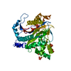 7a12C 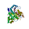 7a13C  7a14C 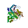 7a15C 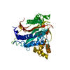 2oazS S: Starting model for refinement C: citing same article ( |
|---|---|
| Similar structure data |


-Links
 PDBj
PDBj


- Assembly
| Deposited unit | 
| ||||||||
|---|---|---|---|---|---|---|---|---|---|
| 1 |
| ||||||||
| Unit cell |
|
-Components
| #1: Protein | Methionyl aminopeptidase / MetAP 2 / Initiation factor 2-associated 67 kDa glycoprotein / p67eIF2 / Peptidase M Mass: 41510.141 Da / Num. of mol.: 1 Source method: isolated from a genetically manipulated source Source: (gene. exp.) Homo sapiens (human) / Gene: METAP2, MNPEP, P67EIF2 / Production host:  unidentified baculovirus / References: UniProt: P50579, methionyl aminopeptidase unidentified baculovirus / References: UniProt: P50579, methionyl aminopeptidase | ||||||||
|---|---|---|---|---|---|---|---|---|---|
| #2: Chemical | ChemComp-PO4 / Phosphate  Mass: 94.971 Da / Num. of mol.: 7 / Source method: obtained synthetically / Formula: PO4 Mass: 94.971 Da / Num. of mol.: 7 / Source method: obtained synthetically / Formula: PO4#3: Chemical | ChemComp-HZE / | 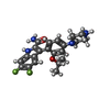  Mass: 414.448 Da / Num. of mol.: 1 / Source method: obtained synthetically / Formula: C22H24F2N4O2 / Feature type: SUBJECT OF INVESTIGATION Mass: 414.448 Da / Num. of mol.: 1 / Source method: obtained synthetically / Formula: C22H24F2N4O2 / Feature type: SUBJECT OF INVESTIGATION#4: Chemical |   Mass: 54.938 Da / Num. of mol.: 2 / Source method: obtained synthetically / Formula: Mn Mass: 54.938 Da / Num. of mol.: 2 / Source method: obtained synthetically / Formula: Mn#5: Water | ChemComp-HOH / | Water Mass: 18.015 Da / Num. of mol.: 395 / Source method: isolated from a natural source / Formula: H2O Mass: 18.015 Da / Num. of mol.: 395 / Source method: isolated from a natural source / Formula: H2OHas ligand of interest | Y | |
-Experimental details
-Experiment
| Experiment | Method: X-RAY DIFFRACTION / Number of used crystals: 1 |
|---|
- Sample preparation
| Crystal | Density Matthews: 2.72 Å3/Da / Density % sol: 54.78 % |
|---|---|
| Crystal grow | Temperature: 293 K / Method: vapor diffusion, hanging drop / pH: 7.4 Details: 200mM potassium phosphate pH 7.4, 100mM MnCl2, 5mM dithiothreitol (DTT) and 25-50% 2-methyl-2,4-pentanediol (MPD) |
-Data collection
| Diffraction | Mean temperature: 100 K / Serial crystal experiment: N |
|---|---|
| Diffraction source | Source: ROTATING ANODE / Type: RIGAKU MICROMAX-007 HF / Wavelength: 1.54 Å |
| Detector | Type: MAR scanner 345 mm plate / Detector: IMAGE PLATE / Date: Nov 17, 2008 |
| Radiation | Protocol: SINGLE WAVELENGTH / Monochromatic (M) / Laue (L): M / Scattering type: x-ray |
| Radiation wavelength | Wavelength: 1.54 Å / Relative weight: 1 |
| Reflection | Resolution: 1.9→26.32 Å / Num. all: 159346 / Num. obs: 35729 / % possible obs: 99.04 % / Redundancy: 4.5 % / Biso Wilson estimate: 23.5 Å2 / Rmerge(I) obs: 0.065 / Net I/σ(I): 12 |
| Reflection shell | Resolution: 1.9→1.968 Å / Redundancy: 2.6 % / Rmerge(I) obs: 0.176 / Mean I/σ(I) obs: 3.3 / Num. unique obs: 15917 / % possible all: 94.53 |
- Processing
| Software |
| ||||||||||||||||||||||||||||||||||||||||||||||||||||||||||||||||||||||||||||||||||||||||||||||||||||||||||||
|---|---|---|---|---|---|---|---|---|---|---|---|---|---|---|---|---|---|---|---|---|---|---|---|---|---|---|---|---|---|---|---|---|---|---|---|---|---|---|---|---|---|---|---|---|---|---|---|---|---|---|---|---|---|---|---|---|---|---|---|---|---|---|---|---|---|---|---|---|---|---|---|---|---|---|---|---|---|---|---|---|---|---|---|---|---|---|---|---|---|---|---|---|---|---|---|---|---|---|---|---|---|---|---|---|---|---|---|---|---|
| Refinement | Method to determine structure: MOLECULAR REPLACEMENT Starting model: 2OAZ Resolution: 1.9→26.32 Å / Cor.coef. Fo:Fc: 0.932 / Cor.coef. Fo:Fc free: 0.916 / SU R Cruickshank DPI: 0.137 / Cross valid method: THROUGHOUT / σ(F): 0 / SU R Blow DPI: 0.151 / SU Rfree Blow DPI: 0.132 / SU Rfree Cruickshank DPI: 0.125
| ||||||||||||||||||||||||||||||||||||||||||||||||||||||||||||||||||||||||||||||||||||||||||||||||||||||||||||
| Displacement parameters | Biso max: 123.5 Å2 / Biso mean: 31.96 Å2 / Biso min: 13.66 Å2
| ||||||||||||||||||||||||||||||||||||||||||||||||||||||||||||||||||||||||||||||||||||||||||||||||||||||||||||
| Refine analyze | Luzzati coordinate error obs: 0.24 Å | ||||||||||||||||||||||||||||||||||||||||||||||||||||||||||||||||||||||||||||||||||||||||||||||||||||||||||||
| Refinement step | Cycle: final / Resolution: 1.9→26.32 Å
| ||||||||||||||||||||||||||||||||||||||||||||||||||||||||||||||||||||||||||||||||||||||||||||||||||||||||||||
| Refine LS restraints |
| ||||||||||||||||||||||||||||||||||||||||||||||||||||||||||||||||||||||||||||||||||||||||||||||||||||||||||||
| LS refinement shell | Resolution: 1.9→1.96 Å / Rfactor Rfree error: 0 / Total num. of bins used: 18
|
